Mastoid cells
| Mastoid cells. | |
|---|---|
 Coronal section of right temporal bone. (Mastoid cells labeled at bottom left.) | |
| Details | |
| Artery | stylomastoid artery |
| Identifiers | |
| Latin | cellulae mastoideae |
| TA | A15.3.02.029 |
| FMA | 72001 |

A section of the mastoid process of the temporal bone of the cranium shows it to be hollowed out into a number of spaces, the mastoid cells, which exhibit great variety in their size and number.
At the upper and front part of the process they are large and irregular and contain air (a form of skeletal pneumaticity), but toward the lower part they diminish in size, while those at the apex of the process are frequently quite small and contain marrow; occasionally they are entirely absent, and the mastoid is then solid throughout.
At birth the mastoid is not pneumatized, but becomes aerated over the first year of life. Poor pneumatization is associated with eustachian tube dysfunction.
Clinical significance
Infections in the middle ear can easily spread into the mastoid area via the aditus ad antrum and mastoid antrum.
Additional images
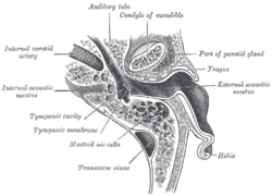 Horizontal section through left ear; upper half of section.
 Mastoid cells.
 Lateral head anatomy detail.Facial nerve dissection.
References
This article incorporates text in the public domain from the 20th edition of Gray's Anatomy (1918)
External links
| Wikimedia Commons has media related to Mastoid cells. |
- Anatomy image: hn1-8 at the College of Medicine at SUNY Upstate Medical University