Mastoid foramen
| Mastoid foramen | |
|---|---|
 Left temporal bone. Inner surface. (Mastoid foramen labeled at bottom left.) | |
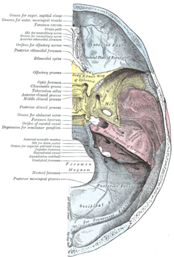 Base of the skull. Upper surface. (Temporal bone is pink, and label for mastoid foramen is at left, second from the bottom.) | |
| Details | |
| Identifiers | |
| Latin | foramen mastoideum |
| TA | A02.1.06.008 |
| FMA | 53159 |
The mastoid foramen is a large hole in the posterior border of the temporal bone. It transmits a Mastoid emissary vein to the sigmoid sinus and a small branch of the occipital artery, the posterior meningeal artery to the dura mater.
Variations
The position and size of this foramen are very variable; it is not always present; sometimes it is situated in the occipital bone, or in the suture between the temporal and the occipital.
It transmits (1) an emissary vein connecting the sigmoid sinus with the posterior auricular vein and (2) a meningeal branch of the occipital artery
Additional images
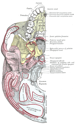 Base of skull. Inferior surface.
References
This article incorporates text in the public domain from the 20th edition of Gray's Anatomy (1918)
External links
- Anatomy diagram: 34257.000-1 at Roche Lexicon - illustrated navigator, Elsevier
- Anatomy diagram: 34257.000-2 at Roche Lexicon - illustrated navigator, Elsevier
- Akram Abood Jaffar: Personal website, Anatomical variations
This article is issued from Wikipedia - version of the 6/9/2015. The text is available under the Creative Commons Attribution/Share Alike but additional terms may apply for the media files.