Frontal sinus
| Frontal sinus | |
|---|---|
 | |
 Nose and nasal cavities | |
| Details | |
| Artery | supra-orbital, anterior ethmoidal |
| Nerve | supraorbital nerve |
| Identifiers | |
| Latin | sinus frontales |
| MeSH | A04.531.621.387 |
| TA | A06.1.03.004 |
| FMA | 57417 |
The frontal sinuses are situated behind the brow ridges. Sinuses are mucosa-lined airspaces within the bones of the face and skull. Each opens into the anterior part of the corresponding middle nasal meatus of the nose through the frontonasal duct which traverses the anterior part of the labyrinth of the ethmoid. These structures then open into the hiatus semilunaris in the middle meatus.
Structure
Frontal sinuses are rarely symmetrical and the septum between them frequently deviates to one or other side of the middle line. Their average measurements are as follows: height 28 mm, breadth 24 mm, depth 20 mm, creating a space of 6-7 ml.[1]
The mucous membrane in this sinus is innervated by the supraorbital nerve, which carries the postganglionic parasympathetic nerve fibers for mucous secretion from the ophthalmic nerve and supplied by the supraorbital artery and anterior ethmoidal artery.
Development
The frontal sinuses are absent at birth, but are generally fairly well developed between the seventh and eighth years, only reaching their full size after puberty.[2] The frontal bone is membranous at birth and there is rarely more than a recess until the bone tissue starts to ossify about age two. Consequently this structure does not show on radiographs before that time. Sinus development begins in the womb, but only the maxillary and ethmoid sinuses are present at birth. Approximately 5% of people have absent frontal sinuses.[3]
Function
Through its copious mucus production, the sinus is an essential part of the immune defense/air filtration carried out by the nose. Nasal and sinal mucosae are ciliated and move mucus to the choanae and finally to the stomach. The thick upper layers of nasal mucus trap bacteria and small particles in tissue abundantly provided with immune cells, antibodies, and antibacterial proteins. The layers beneath are thinner and provide a substrate in which the cilia are able to beat and move the upper layer with its debris through the ostia toward the choanae.
Clinical significance
Infection of the frontal sinus causing sinusitis can give rise to serious complications, as it is in close proximity to the orbit and cranial cavity (orbital cellulitis, epidural and subdural abscess, meningitis).[2]
Fractures
Frontal sinus fractures occur from trauma to the part of the frontal bone that overlies the sinus, often from motor vehicle accidents and falls. The hallmarks of a frontal sinus fracture is a frontal depression in the anterior table of the bone. Additionally, clear fluid leaking from the nose may indicate that fractures to the posterior table have torn into the dura mater, creating a cerebrospinal fluid leak.[4]
Goals in management are to protect the intracranial structure, control any existing CSF leakage, prevent late complications, and aesthetically correct the deformity caused, if any. In anterior table fractures, if the table is minimally displaced, there will be no treatment necessary, only observation. If largely displaced, the correction is open reduction and internal fixation. If inhibiting the nasofrontal outflow tract, procedure is to undergo open reduction and internal fixation of the anterior table and osteoplastic flap with obliteration.
In posterior table fractures, a nondiplaced facture with no CSF leak will only be observed. Those with a CSF leak will undergo sinus exploration if the CSF leak is not internally resolved within 4 to 7 days. With more dramatic displacements, sinus exploration will be required to determine the required level of cranialization, obliteration, and reparation to the dura.
Additional images
 Frontal sinus
 Frontal bone. Inner surface.
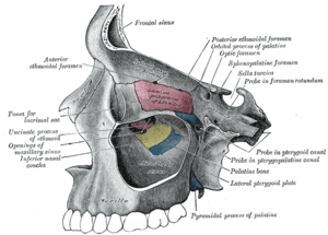 Medial wall of left orbit.
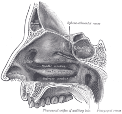 Lateral wall of nasal cavity.
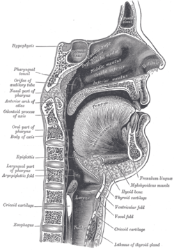 Sagittal section of nose mouth, pharynx, and larynx.
 Outline of bones of face, showing position of air sinuses.
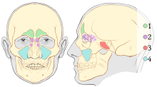 Paranasal sinuses
 Nose diagram
 Frontal sinus
See also
References
This article incorporates text in the public domain from the 20th edition of Gray's Anatomy (1918)
- ↑ University of Texas Medical Branch
- 1 2 Human Anatomy, Jacobs, Elsevier, 2008, page 210
- ↑ The University of Texas Medical Branch, Department of Otolaryngology/Head and Neck Surgery
- ↑ Echo, Anthony; Troy, Jared; Hollier, Larry (29 December 2010). "Frontal Sinus Fractures". Seminars in Plastic Surgery. 24 (04): 375–382. doi:10.1055/s-0030-1269766.
External links
| Wikimedia Commons has media related to Frontal sinuses. |
- Anatomy photo:33:st-0703 at the SUNY Downstate Medical Center
- lesson9 at The Anatomy Lesson by Wesley Norman (Georgetown University) (latnasalwall3, nasalcavitfrontsec)