Stapes
| Stapes, Stirrup | |
|---|---|
Stapes
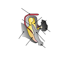
Bones and muscles in the tympanic cavity in the middle ear | |
 Frontal view of stapes (A), and view from below (B). | |
| Details | |
| Precursor | 2nd branchial arch |
| Articulations | Incudostapedial joint |
| Identifiers | |
| Latin | Stapes |
| MeSH | A09.246.397.247.806 |
| TA | A15.3.02.033 |
| FMA | 52751 |
 |
| This article is one of a series documenting the anatomy of the |
| Human ear |
|---|
|
The stapes /ˈsteɪpiːz/ or stirrup is a bone in the middle ear of humans and other mammals which is involved in the conduction of sound vibrations to the inner ear. The stirrup-shaped small bone is on and transmits these to the oval window, medially. The stapes is the smallest and lightest named bone in the human body, and is so-called because of its resemblance to a stirrup (Latin: Stapes).
Structure
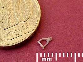
The stapes is the third bone of the three ossicles in the middle ear. The stapes is a stirrup-shaped bone, and the smallest in the human body. It rests on the oval window, to which it is connected by an annular ligament. The stapes is described as having a base, resting on the oval window, as well as a head that articulates with the incus. These are connected by anterior and posterior limbs (Latin: crura).[1]:862 The stapes articulates with the incus through the incudostapedial joint.[2] The stapes is the smallest bone in the human body, and measures roughly 3 x 2.5mm, greater along the head-base span.[3]
Development
The stapes develops from the second pharyngeal arch during the sixth to eighth week of embryological life. The central cavity of the stapedius is due to the presence embryologically of the stapedial artery, which later regresses.[2][4]
Animals
The stapes is one of three ossicles in mammals. In non-mammalian four-legged animals, the bone homologous to the stapes is usually called the columella; however, in reptiles, either term may be used. In fish, the homologous bone is called the hyomandibular, and is part of the gill arch supporting either the spiracle or the jaw, depending on the species. The equivalent term in amphibians is the pars media plectra.[2][5]:481–482
Variation
The stapes appears to be relatively constant in size in different ethnic groups.[6] In 0.01-0.02% of people, the stapedial artery does not regress, and persists in the central foramen.[7] In this case, a pulsatile sound may be heard in the affected ear, or there may be no symptoms at all.[8] Rarely, the stapes may be completely absent.[9][10]:262
Function
Situated between the incus and the inner ear, the stapes transmits sound vibrations from the incus to the oval window, a membrane-covered opening to the inner ear. The stapes is also stabilized by the stapedius muscle, which is innervated by the facial nerve.[1]:861–863
Clinical relevance
Otosclerosis is a congenital or spontaneous-onset disease characterized by abnormal bone remodeling in the inner ear. Often this causes the stapes to adhere to the oval window, which impedes its ability to conduct sound, and is a cause of conductive hearing loss. Clinical otosclerosis is found in about 1% of people, although it is more common in forms that do not cause noticeable hearing loss. Otosclerosis is more likely in young age groups, and females.[11] Two common treatments are stapedectomy, the surgical removal of the stapes and replacement with an artificial prosthesis, and stapedotomy, the creation of a small hole in the base of the stapes followed by the insertion of an artificial prosthesis into that hole.[12] :661 Surgery may be complicated by a persistent stapedial artery, fibrosis-related damage to the base of the bone, or obliterative otosclerosis, resulting in obliteration of the base.[7][10] :254–262
History

The stapes is commonly described as having been discovered by the professor Giovanni Filippo Ingrassia in 1546 at the University of Naples,[13] although this remains the nature of some controversy, as Ingrassia's description was published posthumously in his 1603 anatomical commentary In Galeni librum de ossibus doctissima et expectatissima commentaria. Spanish anatomist Pedro Jimeno is first to have been credited with a published description, in Dialogus de re medica (1549).[14] The bone is so-named because of its resemblance to a stirrup (Latin: stapes), an example of a late Latin word, probably created in mediaeval times from "to stand" (Latin: stapia), as stirrups did not exist in the early Latin-speaking world.[15]
References
- 1 2 Drake, Richard L.; Vogl, Wayne; Tibbitts, Adam W.M. Mitchell; illustrations by Richard; Richardson, Paul (2005). Gray's anatomy for students. Philadelphia: Elsevier/Churchill Livingstone. ISBN 978-0-8089-2306-0.
- 1 2 3 Chapman, SC (Jan 1, 2011). "Can you hear me now? Understanding vertebrate middle ear development.". Frontiers in Bioscience. 16: 1675–92. doi:10.2741/3813. PMID 21196256.
- ↑ aWengen, DF; Nishihara, S; Kurokawa, H; Goode, RL (April 1995). "Measurements of the stapes superstructure.". The Annals of Otology, Rhinology, and Laryngology. 104 (4 Pt 1): 311–6. PMID 7717624.
- ↑ Rodriguez-Vazquez, J. F. (August 2005). "Development of the stapes and associated structures in human embryos". Journal of Anatomy. 207 (2): 165–173. doi:10.1111/j.1469-7580.2005.00441.x.
- ↑ Romer, Alfred Sherwood; Parsons, Thomas S (1977). The Vertebrate Body. Philadelphia, PA: Holt-Saunders International. ISBN 0-03-910284-X.
- ↑ Arensburg, B.; Harell, M.; Nathan, H. (February 1981). "The human middle ear ossicles: Morphometry, and taxonomic implications". Journal of Human Evolution. 10 (2): 199–205. doi:10.1016/S0047-2484(81)80018-8.
- 1 2 Mutlu, C; da Costa, SS; Paparella, MM; Schachern, PA (1998). "Clinical-histopathological correlations of pitfalls in middle ear surgery.". European Archives of Oto-Rhino-Laryngology. 255 (4): 189–94. doi:10.1007/s004050050041. PMID 9592676. Cite uses deprecated parameter
|coauthors=(help) - ↑ Silbergleit, R; Quint, DJ; Mehta, BA; Patel, SC; Metes, JJ; Noujaim, SE (Mar 2000). "The persistent stapedial artery.". American Journal of Neuroradiology. 21 (3): 572–7. PMID 10730654.
- ↑ REIBER, M; SCHWABER, M (February 1997). "Congenital absence of stapes and facial nerve dehiscence". Otolaryngology - Head and Neck Surgery. 116 (2): 278–278. doi:10.1016/S0194-5998(97)70343-7.
- 1 2 Tympanoplasty, Mastoidectomy, and Stapes Surgery. Georg Thieme Verlag. 2008. ISBN 978-1-282-86537-2.
- ↑ Menger, D.J.; Tange, R.A. (April 2003). "The aetiology of otosclerosis: a review of the literature". Clinical Otolaryngology and Allied Sciences. 28 (2): 112–120. doi:10.1046/j.1365-2273.2003.00675.x.
- ↑ Hall, Arthur C. Guyton, John E. (2005). Textbook of medical physiology (11th ed.). Philadelphia: W.B. Saunders. ISBN 978-0-7216-0240-0.
- ↑ Dispenza, F; Cappello, F; Kulamarva, G; De Stefano, A (October 2013). "The discovery of stapes.". Acta Otorhinolaryngologica Italica. 33 (5): 357–9. PMID 24227905.
- ↑ Mudry, Albert (April 2013). "Disputes Surrounding the Discovery of the Stapes in the Mid 16th Century". Otology & Neurotology. 34 (3): 588–592. doi:10.1097/MAO.0b013e31827d8abc.
- ↑ Harper, Douglas. "Stapes (n.)". Online Etymology Dictionary. Retrieved 27 December 2013.
External links
| Wikimedia Commons has media related to Stapes. |
- "3-D Virtual Models of the Human Temporal Bone and Related Structures". Eaton Peabody Laboratory of Auditory Physiology. Retrieved September 8, 2016.