Subarcuate fossa
| Subarcuate fossa | |
|---|---|
 Left temporal bone. Inner surface. (Subarcuate fossa not labeled, but aquaeductus vestibuli labeled at lower right.) | |
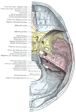 Base of the skull. Upper surface. (Subarcuate fossa not labeled, but temporal bone is identified in pink, and "Eminentia arcuata" is labeled.) | |
| Details | |
| Identifiers | |
| Latin | Fossa subarcuata ossis temporalis |
| TA | A02.1.06.034 |
| FMA | 56418 |
In the temporal bone at the sides of the skull, above and between the aquæductus vestibuli is an irregular depression which lodges a process of the dura mater and transmits a small vein and the subarcuate artery[1] a branch of the meatal segment of anterior inferior cerebellar artery, which is an end artery that supplies blood to the inner ear ; in the infant this depression is represented by a large fossa, the subarcuate fossa, which extends backward as a blind tunnel under the superior semicircular canal.
It is extensive in most primates (except for great apes) and nearly all mammals. In these animals, the subarcuate fossa houses a part of the cerebellum, the petrosal lobe.[2][3]
References
This article incorporates text in the public domain from the 20th edition of Gray's Anatomy (1918)
- ↑ Mom T, Chazal J, Gabrillargues J, Gilain L, Avan P (2005). "Cochlear blood supply: an update on anatomy and function" (PDF). Fr ORL. 88: 81–8.
- ↑ Gannon PJ, Eden AR, Laitman JT (Oct 1988). "The subarcuate fossa and cerebellum of extant primates: comparative study of a skull-brain interface". Am J Phys Anthropol. 77 (2): 143–64. doi:10.1002/ajpa.1330770202. PMID 3207165.
- ↑ Jeffery N, Ryan TM, Spoor F (Aug 2008). "The primate subarcuate fossa and its relationship to the semicircular canals part II: adult interspecific variation". J Hum Evol. 55 (2): 326–39. doi:10.1016/j.jhevol.2008.02.010. PMID 18395770.
External links
- Anatomy diagram: 34257.000-2 at Roche Lexicon - illustrated navigator, Elsevier