Yersinia pestis
| Yersinia pestis | |
|---|---|
 | |
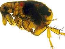
| A scanning electron micrograph depicting a mass of Yersinia pestis bacteria in the foregut of an infected flea and shiot | |
| Scientific classification | |
| Domain: | Bacteria |
| Kingdom: | Eubacteria |
| Phylum: | Proteobacteria |
| Class: | Gammaproteobacteria |
| Order: | Enterobacteriales |
| Family: | Enterobacteriaceae |
| Genus: | Yersinia |
| Species: | Y. pestis |
| Binomial name | |
| Yersinia pestis (Lehmann & Neumann, 1896) van Loghem, 1944 | |
| Synonyms | |
| |
Yersinia pestis[1] (formerly Pasteurella pestis) is a Gram-negative, rod-shaped coccobacillus, a facultative anaerobic organism that can infect humans via the oriental rat flea.[2] It causes the deadly disease called Bubonic Plague (or "the Plague" colloquially).[3][4] Human Y. pestis infection takes three main forms: pneumonic, septicemic, and bubonic plagues.[2] All three forms were responsible for a number of high-mortality epidemics throughout human history, including: the sixth century's Plague of Justinian; the Black Death, which accounted for the death of at least one-third of the European population between 1347 and 1353; and the 19th century's Third Pandemic.[5][6][7][8] These plagues probably originated in China and were transmitted west via trade routes.[8][9]
Y. pestis was discovered in 1894 by Alexandre Yersin, a Swiss/French physician and bacteriologist from the Pasteur Institute, during an epidemic of the plague in Hong Kong.[10] Yersin was a member of the Pasteur school of thought. Kitasato Shibasaburō, a German-trained Japanese bacteriologist who practised Koch's methodology, was also engaged at the time in finding the causative agent of the plague.[11] However, Yersin actually linked plague with Y. pestis. Named Pasteurella pestis in the past, the organism was renamed Yersinia pestis in 1944.
Every year, thousands of cases of the plague are still reported to the World Health Organization, although, with proper treatment, the prognosis for victims is now much better. A five- to six-fold increase in cases occurred in Asia during the time of the Vietnam War, possibly due to the disruption of ecosystems and closer proximity between people and animals. Plague also has a detrimental effect on non-human mammals. In the United States, mammals such as the black-tailed prairie dog and the endangered black-footed ferret are under threat.
General characteristics
Y. pestis is a non-motile, stick-shaped, facultative anaerobic bacterium with bipolar staining (giving it a safety pin appearance) that produces an anti-phagocytic slime layer.[12] Similar to other Yersinia species, it tests negative for urease, lactose fermentation, and indole.[13] The closest relative is the gastrointestinal pathogen Yersinia pseudotuberculosis, and more distantly Yersinia enterocolitica.
Genome
The complete genomic sequence is available for two of the three subspecies of Y. pestis: strain KIM (of biovar Y. p. medievalis),[14] and strain CO92 (of biovar Y. p. orientalis, obtained from a clinical isolate in the United States).[15] As of 2006, the genomic sequence of a strain of biovar Antiqua has been recently completed.[16] Similar to the other pathogenic strains, there are signs of loss of function mutations. The chromosome of strain KIM is 4,600,755 base pairs long; the chromosome of strain CO92 is 4,653,728 base pairs long. Like Y. pseudotuberculosis and Y. enterocolitica, Y. pestis is host to the plasmid pCD1. In addition, it also hosts two other plasmids, pPCP1 (also called pPla or pPst) and pMT1 (also called pFra) that are not carried by the other Yersinia species. pFra codes for a phospholipase D that is important for the ability of Y. pestis to be transmitted by fleas.[17] pPla codes for a protease, Pla, that activates plasmin in human hosts and is a very important virulence factor for pneumonic plague.[18] Together, these plasmids, and a pathogenicity island called HPI, encode several proteins that cause the pathogenesis, for which Y. pestis is famous. Among other things, these virulence factors are required for bacterial adhesion and injection of proteins into the host cell, invasion of bacteria in the host cell (via a type-III secretion system), and acquisition and binding of iron harvested from red blood cells (by siderophores). Y. pestis is thought to be descendant from Y. pseudotuberculosis, differing only in the presence of specific virulence plasmids.
A comprehensive and comparative proteomics analysis of Y. pestis strain KIM was performed in 2006.[19] The analysis focused on the transition to a growth condition mimicking growth in host cells.
small non-coding RNA
Numerous bacterial small non-coding RNAs have been identified to play regulatory functions. Some can regulate the virulence genes. 63 novel putative sRNA were identified through deep sequencing of the Y. pestis sRNA-ome. Among them was Yersinia-specific (also present in Y. pseudotuberculosis and Y. enterocolitica) Ysr141 (Yersinia small RNA 141). Ysr141 sRNA was shown to regulate the synthesis of the type III secretion system (T3SS) effector protein YopJ.[20] The Yop-Ysc T3SS is a critical component of virulence for Yersinia species.[21]
Pathogenesis and immunity
In the urban and sylvatic (forest) cycles of Y. pestis, most of the spreading occurs between rodents and fleas. In the sylvatic cycle, the rodent is wild, but in the urban cycle, the rodent is primarily the brown rat. In addition, Y. pestis can spread from the urban environment and back. Transmission to humans is usually through the bite of infected fleas. If the disease has progressed to the pneumonic form, humans can spread the bacterium to others by coughing, vomiting, and possibly sneezing.
In reservoir hosts
Several species of rodents serve as the main reservoir for Y. pestis in the environment. In the steppes, the natural reservoir is believed to be principally the marmot. In the western United States, several species of rodents are thought to maintain Y. pestis. However, the expected disease dynamics have not been found in any rodent. A variety of species of rodents are known to have a variable resistance, which could lead to an asymptomatic carrier status.[22] Evidence indicates fleas from other mammals have a role in human plague outbreaks.[23]
The lack of knowledge of the dynamics of plague in mammal species is also true among susceptible rodents such as the black-tailed prairie dog (Cynomys ludovicianus), in which plague can cause colony collapse, resulting in a massive effect on prairie food webs.[24] However, the transmission dynamics within prairie dogs does not follow the dynamics of blocked fleas; carcasses, unblocked fleas, or another vector could possibly be important, instead.[25]
In other regions of the world, the reservoir of the infection is not clearly identified, which complicates prevention and early warning programs. One such example was seen in a 2003 outbreak in Algeria.[26] The domestic house cat is susceptible to plague. Their symptoms are similar to those experienced by humans. Cats infected with plague can infect people through bites, scratches, coughs, or sneezes.[27]
Vector
The transmission of Y. pestis by fleas is well characterized.[28] Initial acquisition of Y. pestis by the vector occurs during feeding on an infected animal. Several proteins then contribute to the maintenance of the bacteria in the flea digestive tract, among them the hemin storage system and Yersinia murine toxin (Ymt). Although Ymt is highly toxic to rodents and was once thought to be produced to ensure reinfection of new hosts, it is important for the survival of Y. pestis in fleas.[17]
The hemin storage system plays an important role in the transmission of Y. pestis back to a mammalian host.[29] While in the insect vector, proteins encoded by hemin storage system genetic loci induce biofilm formation in the proventriculus, a valve connecting the midgut to the esophagus.[30] Aggregation in the biofilm inhibits feeding, as a mass of clotted blood and bacteria forms (referred to as "Bacot's block"[31]). Transmission of Y. pestis occurs during the futile attempts of the flea to feed. Ingested blood is pumped into the esophagus, where it dislodges bacteria lodged in the proventriculus and is regurgitated back into the host circulatory system.
In humans and other susceptible hosts
Pathogenesis due to Y. pestis infection of mammalian hosts is due to several factors, including an ability of these bacteria to suppress and avoid normal immune system responses such as phagocytosis and antibody production. Flea bites allow for the bacteria to pass the skin barrier. Y. pestis expresses the yadBC gene, which is similar to adhesins in other Yersinia species, allowing for adherence and invasion of epithelial cells.[32] Y. pestis expresses a plasmin activator that is an important virulence factor for pneumonic plague and that might degrade on blood clots to facilitate systematic invasion.[18] Many of the bacteria's virulence factors are anti-phagocytic in nature. Two important anti-phagocytic antigens, named F1 (Fraction 1) and V or LcrV, are both important for virulence.[12] These antigens are produced by the bacterium at normal human body temperature. Furthermore, Y. pestis survives and produces F1 and V antigens while it is residing within white blood cells such as monocytes, but not in neutrophils. Natural or induced immunity is achieved by the production of specific opsonic antibodies against F1 and V antigens; antibodies against F1 and V induce phagocytosis by neutrophils.[33]
In addition, the type-III secretion system (T3SS) allows Y. pestis to inject proteins into macrophages and other immune cells. These T3SS-injected proteins, called Yersinia outer proteins (Yops), include Yop B/D, which form pores in the host cell membrane and have been linked to cytolysis. The YopO, YopH, YopM, YopT, YopJ, and YopE are injected into the cytoplasm of host cells by T3SS into the pore created in part by YopB and YopD.[34] The injected Yops limit phagocytosis and cell signaling pathways important in the innate immune system, as discussed below. In addition, some Y. pestis strains are capable of interfering with immune signaling (e.g., by preventing the release of some cytokines).
Y. pestis proliferates inside lymph nodes, where it is able to avoid destruction by cells of the immune system such as macrophages. The ability of Y. pestis to inhibit phagocytosis allows it to grow in lymph nodes and cause lymphadenopathy. YopH is a protein tyrosine phosphatase that contributes to the ability of Y. pestis to evade immune system cells.[35] In macrophages, YopH has been shown to dephosphorylate p130Cas, Fyb (Fyn binding protein) SKAP-HOM and Pyk, a tyrosine kinase homologous to FAK. YopH also binds the p85 subunit of phosphoinositide 3-kinase, the Gab1, the Gab2 adapter proteins, and the Vav guanine nucleotide exchange factor.
YopE functions as a GTPase-activating protein for members of the Rho family of GTPases such as RAC1. YopT is a cysteine protease that inhibits RhoA by removing the isoprenyl group, which is important for localizing the protein to the cell membrane. It has been proposed that YopE and YopT may function to limit YopB/D-induced cytolysis.[36] This might limit the function of YopB/D to create the pores used for Yop insertion into host cells and prevent YopB/D-induced rupture of host cells and release of cell contents that would attract and stimulate immune system responses.
YopJ is an acetyltransferase that binds to a conserved α-helix of MAPK kinases.[37] YopJ acetylates MAPK kinases at serines and threonines that are normally phosphorylated during activation of the MAP kinase cascade.[38][39] YopJ is activated in eukaryotic cells by interaction with target cell Phytic acid (IP6).[40] This disruption of host cell protein kinase activity causes apoptosis of macrophages, and it has been proposed that this is important for the establishment of infection and for evasion of the host immune response. YopO is a protein kinase also known as Yersinia protein kinase A (YpkA). YopO is a potent inducer of human macrophage apoptosis.[41]
Depending on which form of the plague the individual becomes infected with the plague develops different illness; however the plague overall effects the hosts cell’s ability to communicate with the immune system, hindering the body to bring phagocytic cells to the area of infection.
Immunity
A formalin-inactivated vaccine once was available in the United States for adults at high risk of contracting the plague until removal from the market by the Food and Drug Administration. It was of limited effectiveness and could cause severe inflammation. Experiments with genetic engineering of a vaccine based on F1 and V antigens are underway and show promise. However, bacteria lacking antigen F1 are still virulent, and the V antigens are sufficiently variable, such that vaccines composed of these antigens may not be fully protective.[42] United States Army Medical Research Institute of Infectious Diseases (USAMRIID) have found that an experimental F1/V antigen-based vaccine protects crab-eating macaques but fails to protect African green monkey species.[43] A systematic review by the Cochrane Collaboration found no studies of sufficient quality to make any statement on the efficacy of the vaccine.[44]
Clinical aspects
Symptoms and disease progression
- Bubonic plague
- Incubation period of 2–6 days, when the bacterium is actively replicating.
- General malaise
- Fever
- Muscle aches
- Gangrene
- Headache and chills occur suddenly at the end of the incubation period
- Extreme weakness
- Swelling of lymph nodes resulting in buboes, the classic sign of bubonic plague. Most flea bites will occur on the legs, so the inguinal nodes are most frequently affected
- Death can occur in less than 2 weeks
- Septicemic plague
- Nausea
- Vomiting
- Diarrhea
- Abdominal pain
- Hypotension
- Hepatosplenomegaly
- Delirium
- Seizures in children
- Shock
- General malaise
- Fever
- Symptoms of bubonic or pneumonic plague are not always present
Note: Patient may die before any symptoms appear
- Pneumonic plague (Spread person to person)
- Fever
- Chills
- Coughing
- Chest pain
- Sneezing
- Rapid breathing
- Dyspnea
- Hemoptysis
- Lethargy
- Hypotension
- Shock
- Symptoms of bubonic or septicemic plague are not always present[45]
- 100% mortality if not treated
If this occurs with the classic buboes, this is considered primary, while secondary occurs after symptoms of bubonic or pneumonic infection. Since the bacteria are blood-borne, several organs can be affected, including the spleen and brain. The diffuse infection can cause an immunologic cascade to occur, leading to disseminated intravascular coagulation (DIC), which in turn results in bleeding and necrotic skin and tissue. Such a disseminated infection increases mortality to 22%.
With the exception of the buboes, the initial symptoms of plague are very similar to many other diseases, making diagnosis difficult.[46]
ICD-9 codes for the diseases caused by Y. pestis:
- 020.0 Bubonic plague
- 020.2 Septicemic plague
- 020.3 Primary pneumonic plague
- 020.4 Secondary pneumonic plague
- 020.5 Unspecified pneumonic plague
Clinical determination
Gram's stains can confirm the presence of gram-negative rods, and in some cases the identification of the double-curved shape. An anti-F1 serology test can differentiate between different species of Yersinia, and Polymerase chain reaction (PCR) can be used to identify Y. pestis.
The protein H of the tail fiber of the bacteriophage Yersinia phage L-413C permits the differentiation between Y. pestis and Y. pseudotuberculosis", the gastro-intestinal corrolary (Kane et al.).[47]
Treatment
The traditional second line treatment for Y. pestis has been streptomycin,[48][49] chloramphenicol, tetracycline,[50] and fluoroquinolones.[51] There is also good evidence to support the use of doxycycline or gentamicin.[52] Resistant strains have been isolated; treatment should be guided by antibiotic sensitivities where available. Antibiotic treatment alone is insufficient for some patients, who may also require circulatory, ventilator, or renal support.
In an emergency department setting, Harrison's Principles of Internal Medicine outlines the following treatment course.[53] Antibiotics within the first 24 hours are very beneficial, with intravenous being preferred in pulmonary or advanced cases. Streptomycin or gentamicin are the first-line drugs, with chloramphenicol for critically ill patients, or rarely for suspected neuro-involvement.
Prevention
- Avoid contact with sick or dead animals, especially rodents.
- Avoid contact with the nests and burrows of rodents, squirrels, chipmunks, and prairie dogs.
- If living in areas where rodent plague occurs, do use monthly topical or oral flea prevention for dogs and cats. Refrain from allowing pets to roam freely.
- If exposure to rodent fleas is anticipated, apply insect repellent containing DEET (diethyltoluamide) to the skin.
- Wear gloves when handling infected or dead animals.
Plague vaccine is available, based on an attenuated type.[54]
Historical outbreaks
Plague of Justinian
During the mid-sixth century, the pandemic known as the Plague of Justinian wiped out roughly one third of the Byzantine Empire's population, creating major military and financial difficulties. Modern historians named this plague incident after the Eastern Roman Emperor Justinian I, who held power in the Byzantine capital of Constantinople at the time of the initial outbreak. The primary years of the plague were 541–542 AD, although plague returned throughout the Mediterranean basin in successive generations, until about 750.
The waves of disease had a major effect on the future course of European history. The plague's social and cultural impact during the Justinian period is comparable to that of the Black Death.
The most commonly accepted cause of the pandemic has been bubonic plague.[55] A genetic study suggests the Plague of Justinian (and others from antiquity) arose from either now-extinct strains of Y. pestis, genetically distinct from the strain that broke out in the 14th-century pandemic, or from pathogens entirely unrelated to bubonic plague.[56][57]
Role in Black Death
In the 1340s, Europe had a severe disease outbreak that began in the southern port cities in Italy and thereafter steadily spread northward, killing millions in its path. It often emptied entire villages of people and created mass hysteria. The death toll was so great, a very religious contingent thought God was punishing man for sin. When it finally reached England in 1348, it wound up destroying more than a third of the country's population. From that time on, the disease would recur repeatedly throughout Europe until the end of the 18th century, with the last outbreak in England occurring in 1666.
In 2000, Didier Raoult and others reported finding Y. pestis DNA by performing a "suicide PCR" on tooth pulp tissue from a 14th-century plague cemetery in Montpellier.[58]
A study by an international team of researchers published in October 2010 confirmed Y. pestis was the cause of the Black Death and later epidemics on the entire European continent over a period of 400 years. The team used ancient DNA and proteins recovered from the bodies of plague victims buried in Hereford in England, in Saint-Laurent-de-la-Cabrerisse in France, and Bergen op Zoom in the Netherlands to identify the pathogen.[59] They found two previously unknown, older strains of Y. pestis that had spread from China by two different routes, rather than the modern Y. p., orientalis and Y. p. medievalis.[60]
Three biovars of Y. pestis were originally thought to correspond to one of the historical pandemics of bubonic plague.[61] Biovar Y. p. antiqua is thought to correspond to the Plague of Justinian; it is not known whether this biovar also corresponds to earlier or smaller epidemics of bubonic plague, or whether these were even truly bubonic plague.[62] Biovar Y. p. mediaevalis was formerly thought to correspond to the Black Death, while Biovar Y. p. orientalis was thought to correspond to the third pandemic and the majority of modern outbreaks of plague. However, calculations of Y. pestis' evolutionary age, found using the number of synonymous single-nucleotide polymorphisms (SNPs) in conjunction with molecular clock rates, date the emergence of the biovars prior to any of the historical epidemics due to the length of time needed to accumulate such mutations.[15] Additional evidence against this hypothesis includes the fact that Y. p. mediaevalis is likely too young to have produced the Black Death due to its recent divergence from Y. p. orientalis.[63]
Use in biological warfare
Y. pestis is potentially one of the first examples of biological warfare in (recorded) history, when in 1347, plague victims were catapulted by the Mongols over the city walls of Caffa, a town currently known as Feodosiya located in Crimea. Infected inhabitants may have fled to Italy, thus spreading the Black Death to Europe, though this is likely only one of a few routes that could have brought the plague from the east.[64]
Y. pestis was used as a biological weapon in World War II, when on October 4, 1940, a Japanese airplane flying over Chushien, Chekiang Province, China, released rice and wheat plus rat fleas carrying Y. pestis. A second plane load was released three weeks later. These actions led to a local plague that killed 121 people.[65]
Proposed use in biological warfare
Leon A. Fox from the U.S. Army Medical Corps had suggested a similar approach in 1933, proposing the possibility of infected rats being dropped from planes.[65] U.S. Army Medical Corps Major Leon Fox published an article in 1933 in the magazine Military Surgeon writing: "Practically insurmountable difficulties prevent the use of biologic agents as effective weapons."[66]
Recent events
In September 2009, the death of Malcolm Casadaban, a molecular genetics professor at the University of Chicago, was linked to his work on a weakened laboratory strain of Y. pestis.[67] Hemochromatosis was hypothesised to be a predisposing factor in Casadaban's death from this attenuated strain used for research.[68]
In 2012, researchers in Germany collected samples of Yersinia pestis from gravesites with a view to reconstructing the DNA of the bacterium.[69] In 2015, Cell published results from a study of ancient graves. Plasmids of Y. pestis were detected in archaeological samples of the teeth of seven Bronze Age individuals, in the Afanasievo culture in Siberia, the Corded Ware culture in Estonia, the Sintashta culture in Russia, the Unetice culture in Poland and the Andronovo culture in Siberia.[70]
June 8, 2015 Larimer County, CO Fatality confirmed By CDC as listed on the RSOE EDIS - Emergency and Disaster Information Service[3]
September 8, 2016 Yersinia pestis bacterium identified from DNA in teeth from skeletons found at Crossrail Site, London showing the human remains were victims of the Great Plague http://www.crossrail.co.uk/news/articles/dna-of-bacteria-responsible-for-london-great-plague-of-1665-identified-for-first-time
References
- ↑ Sutyak, Katya. "Yersinia Pestis." University of Connecticut. University of Connecticut, n.d. Web. 10 Nov. 2015.
- 1 2 Ryan KJ, Ray CG, eds. (2004). Sherris Medical Microbiology (4th ed.). McGraw Hill. pp. 484–488. ISBN 0-8385-8529-9.
- 1 2 http://hisz.rsoe.hu/alertmap/site/index.php?pageid=event_desc&edis_id=BH-20150621-48754-USA
- ↑ CDC http://www.cdc.gov/plague/
- ↑ Austin Alchon, Suzanne (2003). A pest in the land: new world epidemics in a global perspective. University of New Mexico Press. p. 21. ISBN 0-8263-2871-7.
- ↑ Harbeck, Michaela; Seifert, Lisa; Hänsch, Stephanie; Wagner, David M.; Birdsell, Dawn; Parise, Katy L.; Wiechmann, Ingrid; Grupe, Gisela; Thomas, Astrid; Keim, Paul; Zöller, Lothar; Bramanti, Barbara; Riehm, Julia M.; Scholz, Holger C. (2013). "Yersinia pestis DNA from Skeletal Remains from the 6th Century AD Reveals Insights into Justinianic Plague". PLoS Pathogens. 9 (5): e1003349. doi:10.1371/journal.ppat.1003349. PMC 3642051
 . PMID 23658525. Lay summary – ScienceDaily (May 10, 2013).
 - ↑ Carter, Adam (Jan 27, 2014). "Black Death mysteries unlocked by McMaster scientists". CBC News.
- 1 2 Nicholas Wade (October 31, 2010). "Europe's Plagues Came From China, Study Finds". New York Times. Retrieved November 1, 2010.
- ↑ Morelli, G.; Song, Y.; Mazzoni, C.J.; Eppinger, M.; Roumagnac, P.; Wagner, D.M.; Feldkamp, M.; Kusecek, B.; Vogler, A.J.; Li, Y.; Cui, Y.; Thomson, N.R.; Jombart, T.; Leblois, R.; Lichtner, P.; Rahalison, L.; Petersen, J.M.; Balloux, F.; Keim, Pl; Wirth, T.; Ravel, J.; Yang, R.; Carniel, E.; Achtman, M. (December 2010). "Yersinia pestis genome sequencing identifies patterns of global phylogenetic diversity". Nature Genetics. 42 (12): 1140–1143. doi:10.1038/ng.705. PMC 2999892. PMID 21037571.
- ↑ Bockemühl J (1994). "100 years after the discovery of the plague-causing agent--importance and veneration of Alexandre Yersin in Vietnam today". Immun Infekt. 22 (2): 72–5. PMID 7959865.
- ↑ Howard-Jones N (1973). "Was Kitasato Shibasaburō the discoverer of the plague bacillus?". Perspect Biol Med. 16 (2): 292–307. PMID 4570035.
- 1 2 Collins FM (1996). Baron S; et al., eds. Pasteurella, Yersinia, and Francisella. In: Baron's Medical Microbiology (4th ed.). Univ. of Texas Medical Branch. ISBN 0-9631172-1-1.
- ↑ Stackebrandt, Erko; Dworkin, Martin; Falkow, Stanley; Rosenberg, Eugene; Karl-Heinz Schleifer (2005). The Prokaryotes: A Handbook on the Biology of Bacteria:Volume 6: Proteobacteria: Gamma Subclass. Berlin: Springer. ISBN 0-387-25499-4.
- ↑ Deng, W.; Burland, V.; Plunkett III, G.; Boutin, A.; Mayhew, G.F.; Liss, P.; Perna, N.T.; Rose, D.J.; Mau, B.; Zhou, S.; Schwartz, D.C.; Fetherston, J.D.; Lindler, L.E.; Brubaker, R.R.; Plano, G.V.; Straley, S.C.; McDonough, K.A.; Nilles, M.L.; Matson, J.S.; Blattner, F.R.; Perry, R.D. (August 2002). "Genome sequence of Yersinia pestis KIM". Journal of Bacteriology. 184 (16): 4601–4611. doi:10.1128/JB.184.16.4601-4611.2002. PMC 135232. PMID 12142430.
- 1 2 Parkhill, J.; Wren, B.W.; Thomson, N.R.; Titball, R.W.; Holden, H.T.; Prentice, M.B.; Sebaihia, M.; James, K.D.; Churcher, C.; Mungall, K.L.; Baker, S.; Basham, D.; Bentley, S.D.; Brooks, K.; Cerdeño-Tárraga, A.M.; Chillingworth, T.; Cronin, A.; Davies, R.M.; Davis, P.; Dougan, G.; Feltwell, T.; Hamlin, N.; Holroyd, S.; Jagels, K.; Karlyshev, A.V.; Leather, S.; Moule, S.; Oyston, P.C.; Quail, M.; Rutherford, K.; Simmonds, M.; Skelton, J.; Stevens, K.; Whitehead, S.; Barrell, B.G. (October 2001). "Genome sequence of Yersinia pestis, the causative agent of plague". Nature. 413 (6855): 523–527. doi:10.1038/35097083. PMID 11586360.
- ↑ Chain PS, Hu P, Malfatti SA, et al. (2006). "Complete Genome Sequence of Yersinia pestis Strains Antiqua and Nepal516: Evidence of Gene Reduction in an Emerging Pathogen". J. Bacteriol. 188 (12): 4453–63. doi:10.1128/JB.00124-06. PMC 1482938. PMID 16740952.
- 1 2 Hinnebusch BJ, Rudolph AE, Cherepanov P, Dixon JE, Schwan TG, Forsberg A (2002). "Role of Yersinia murine toxin in survival of Yersinia pestis in the midgut of the flea vector". Science. 296 (5568): 733–5. Bibcode:2002Sci...296..733H. doi:10.1126/science.1069972. PMID 11976454.
- 1 2 Lathem WW, Price PA, Miller VL, Goldman WE (2007). "A plasminogen-activating protease specifically controls the development of primary pneumonic plague". Science. 315 (5811): 509–13. Bibcode:2007Sci...315..509L. doi:10.1126/science.1137195. PMID 17255510.
- ↑ Hixson, K.K.; Adkins, J.N.; Baker, S.E.; Moore, R.J.; Chromy, B.A.; Smith, R.D.; McCutchen-Maloney, S.L.; Lipton, M.S. (November 2006). "Biomarker candidate identification in Yersinia pestis using organism-wide semiquantitative proteomics". Journal of Proteome Research. 5 (11): 3008–3017. doi:10.1021/pr060179y. PMID 17081052.
- ↑ Schiano, Chelsea A.; Koo, Jovanka T.; Schipma, Matthew J.; Caulfield, Adam J.; Jafari, Nadereh; Lathem, Wyndham W. (2014-05-01). "Genome-wide analysis of small RNAs expressed by Yersinia pestis identifies a regulator of the Yop-Ysc type III secretion system". Journal of Bacteriology. 196 (9): 1659–1670. doi:10.1128/JB.01456-13. ISSN 1098-5530. PMC 3993326. PMID 24532772.
- ↑ Cornelis, G. R.; Boland, A.; Boyd, A. P.; Geuijen, C.; Iriarte, M.; Neyt, C.; Sory, M. P.; Stainier, I. (1998-12-01). "The virulence plasmid of Yersinia, an antihost genome". Microbiology and molecular biology reviews: MMBR. 62 (4): 1315–1352. ISSN 1092-2172. PMC 98948. PMID 9841674.
- ↑ Meyer K.F. (1957). "The natural history of plague and psittacosis: The R. E. Dyer Lecture". Public Health Reports. 72 (8): 705–19. doi:10.2307/4589874. JSTOR 4589874. PMC 2031327. PMID 13453634.
- ↑ von Reyn CF, Weber NS, Tempest B, et al. (1977). "Epidemiologic and clinical features of an outbreak of bubonic plague in New Mexico". J. Infect. Dis. 136 (4): 489–94. doi:10.1093/infdis/136.4.489. PMID 908848.
- ↑ Pauli JN, Buskirk SW, Williams ES, Edwards WH (2006). "A plague epizootic in the black-tailed prairie dog (Cynomys ludovicianus)". J. Wildl. Dis. 42 (1): 74–80. doi:10.7589/0090-3558-42.1.74. PMID 16699150.
- ↑ Webb CT, Brooks CP, Gage KL, Antolin MF (2006). "Classic flea-borne transmission does not drive plague epizootics in prairie dogs". Proc. Natl. Acad. Sci. U.S.A. 103 (16): 6236–41. Bibcode:2006PNAS..103.6236W. doi:10.1073/pnas.0510090103. PMC 1434514. PMID 16603630.
- ↑ Bertherat, E.; Bekhoucha, S.; Chougrani, S.; Razik, F.; Duchemin, J.B.; Houti, L.; Deharib, L.; Fayolle, C.; Makrerougrass, B.; Dali-Yahia, R.; Bellal, R.; Belhabri, L.; Chaieb, A.; Tikhomirov, E.; Carniel, E. (October 2007). "Plague reappearance in Algeria after 50 years, 2003". Emerging Infectious Diseases. 13 (10): 1459–1462. doi:10.3201/eid1310.070284. PMC 2851531. PMID 18257987.
- ↑ "Cats - Healthy Pets Healthy People". Centers for Disease Control and Prevention. 2016-05-13. Retrieved 2016-11-25.
- ↑ Zhou D, Han Y, Yang R (2006). "Molecular and physiological insights into plague transmission, virulence and etiology". Microbes Infect. 8 (1): 273–84. doi:10.1016/j.micinf.2005.06.006. PMID 16182593.
- ↑ B.J. Hinnebusch; R.D. Perry & T.G. Schwan (1996). "Role of the Yersinia pestis hemin storage (hms) locus in the transmission of plague by fleas". Science. 273 (5237): 367–70. Bibcode:1996Sci...273..367H. doi:10.1126/science.273.5273.367. PMID 8662526.
- ↑ Erickson, D. L.; N. R. Waterfield; V. Vadyvaloo; D. Long; E. R. Fischer; R. Ffrench-Constant & B. J. Hinnebusch (2007). "Acute oral toxicity of Yersinia pseudotuberculosis to fleas: Implications for the evolution of vector-borne transmission of plague". Cellular Microbiology. 9 (11): 2658–2666. doi:10.1111/j.1462-5822.2007.00986.x. PMID 17587333.
- ↑ Pepper, C., M. Nascarella, E. Marsland, J. Montford, L. Wood, S. Cox, C. Bradford, T. Burns, and S. Presley. 2004. Threatened or endangered? Keystone species or public health threat? The black-tailed prairie dog, the Endangered Species Act, and the imminent threat of bubonic plague. Journal of Land, Resources, and Environmental Law 24: 355-391.
- ↑ Forman S, Wulff CR, Myers-Morales T, Cowan C, Perry RD, Straley SC (2008). "yadBC of Yersinia pestis, a New Virulence Determinant for Bubonic Plague". Infect. Immun. 76 (2): 578–87. doi:10.1128/IAI.00219-07. PMC 2223446. PMID 18025093.
- ↑ Salyers AA, Whitt DD (2002). Bacterial Pathogenesis: A Molecular Approach (2nd ed.). ASM Press. pp. 207-12.
- ↑ Viboud GI, Bliska JB (2005). "Yersinia outer proteins: role in modulation of host cell signaling responses and pathogenesis". Annu. Rev. Microbiol. 59 (1): 69–89. doi:10.1146/annurev.micro.59.030804.121320. PMID 15847602.
- ↑ de la Puerta ML, Trinidad AG, del Carmen Rodríguez M, Bogetz J, Sánchez Crespo M, Mustelin T, Alonso A, Bayón Y (February 2009). Bozza P, ed. "Characterization of New Substrates Targeted By Yersinia Tyrosine Phosphatase YopH". PLoS ONE. 4 (2): e4431. Bibcode:2009PLoSO...4.4431D. doi:10.1371/journal.pone.0004431. PMC 2637541. PMID 19221593.
- ↑ Mejía E, Bliska JB, Viboud GI (February 2009). "Yersinia Controls Type III Effector Delivery into Host Cells by Modulating Rho Activity". PLoS ONE. 4 (2): e4431. doi:10.1371/journal.ppat.0040003. PMC 2186360. PMID 18193942.
- ↑ Hao YH, Wang Y, Burdette D, Mukherjee S, Keitany G, Goldsmith E, Orth K (January 2008). Kobe B, ed. "Structural Requirements for Yersinia YopJ Inhibition of MAP Kinase Pathways". PLoS ONE. 2 (3): e1375. Bibcode:2008PLoSO...3.1375H. doi:10.1371/journal.pone.0001375. PMC 2147050. PMID 18167536.
- ↑ Mukherjee, S.; Keitany, Gladys; Li, Yan; Wang, Yong; Ball, Haydn L.; Goldsmith, Elizabeth J.; Orth, Kim (2006). "Yersinia YopJ Acetylates and Inhibits Kinase Activation by Blocking Phosphorylation". Science. 312 (5777): 1211–4. Bibcode:2006Sci...312.1211M. doi:10.1126/science.1126867. PMID 16728640.
- ↑ Mittal, R.; Peak-Chew, S.-Y.; McMahon, H. T. (2006). "Acetylation of MEK2 and I B kinase (IKK) activation loop residues by YopJ inhibits signaling". Proceedings of the National Academy of Sciences. 103 (49): 18574–18579. Bibcode:2006PNAS..10318574M. doi:10.1073/pnas.0608995103. PMC 1654131. PMID 17116858.
- ↑ Mittal R, Peak-Chew SY, Sade RS, Vallis Y, McMahon HT (2010). "The Acetyltransferase Activity of the Bacterial Toxin YopJ of Yersinia Is Activated by Eukaryotic Host Cell Inositol Hexakisphosphate". J Biol Chem. 285 (26): 19927–34. doi:10.1074/jbc.M110.126581. PMC 2888404. PMID 20430892.
- ↑ Park H, Teja K, O'Shea JJ, Siegel RM (May 2007). "The Yersinia effector protein YpkA induces apoptosis independently of actin depolymerization". J Immunol. 178 (10): 6426–6434. doi:10.4049/jimmunol.178.10.6426. PMID 17475872.
- ↑ Welkos S, et al. (2002). "Determination of the virulence of the pigmentation-deficient and pigmentation-/plasminogen activator-deficient strains of Yersinia pestis in non-human primate and mouse models of pneumonic plague". Vaccine. 20 (17–18): 2206–2214. doi:10.1016/S0264-410X(02)00119-6. PMID 12009274.
- ↑ Pitt ML (October 2004). "Non-human primates as a model for pneumonic plague. In: Animals Models and Correlates of Protection for Plague Vaccines Workshop" (PDF).
- ↑ Jefferson T, Demicheli V, Pratt M (2000). Jefferson T, ed. "Vaccines for preventing plague". Cochrane Database Syst Rev (2): CD000976. doi:10.1002/14651858.CD000976. PMID 10796565.
- ↑ Info taken from "Harrison's Principles of Internal Medicine 16th Edition"
- ↑ Prentice, Michael B; Rahalison, Lila (2007). "Plague". The Lancet. 369 (9568): 1196–1207. doi:10.1016/S0140-6736(07)60566-2. PMID 17416264.
- ↑ Garcia, E; et al. (2008). "Molecular characterization of L-413C, a P2-related plague diagnostic bacteriophage". Virology. 372 (1): 85–96. doi:10.1016/j.virol.2007.10.032. PMID 18045639.
- ↑ Wagle PM. (1948). "Recent advances in the treatment of bubonic plague". Indian J Med Sci. 2: 489–94.
- ↑ Meyer KF. (1950). "Modern therapy of plague". JAMA. 144 (12): 982–5. doi:10.1001/jama.1950.02920120006003. PMID 14774219.
- ↑ Kilonzo BS, Makundi RH, Mbise TJ (1992). "A decade of plague epidemiology and control in the Western Usambara mountains, north-east Tanzania". Acta Tropica. 50 (4): 323–9. doi:10.1016/0001-706X(92)90067-8. PMID 1356303.
- ↑ WHO/CDS/CSR/EDC/99.2 Plague Manual | World Health Organization | Epidemiology, Distribution, Surveillance and Control | Pages: 1-5
- ↑ Mwengee W, Butler T, Mgema S, et al. (2006). "Treatment of plague with gentamicin or doxycycline in a randomized clinical trial in Tanzania". Clin Infect Dis. 42 (5): 614–21. doi:10.1086/500137. PMID 16447105.
- ↑ Jameson, J. N. St C.; Dennis L. Kasper; Harrison, Tinsley Randolph; Braunwald, Eugene; Fauci, Anthony S.; Hauser, Stephen L & Longo, Dan L. (2005). Harrison's principles of internal medicine. New York: McGraw-Hill Medical Publishing Division. ISBN 0-07-140235-7.
- ↑ Bubeck SS, Dube PH (September 2007). "Yersinia pestis CO92ΔyopH Is a Potent Live, Attenuated Plague Vaccine". Clin. Vaccine Immunol. 14 (9): 1235–8. doi:10.1128/CVI.00137-07. PMC 2043315. PMID 17652523.
- ↑ "Europe's Plagues Came From China, Study Finds". The New York Times. October 31, 2010. Retrieved 2010-11-01.
- ↑ McGrath, Matt (12 October 2011). "Black Death Genetic Code 'Built'". BBC World Service. Retrieved 12 October 2011.
- ↑ Bos, Kirsten; Schuenemann, Verena J.; Golding, G. Brian; Burbano, Hernán A.; Waglechner, Nicholas; Coombes, Brian K.; McPhee, Joseph B.; Dewitte, Sharon N.; Meyer, Matthias; Schmedes, Sarah; Wood, James; Earn, David J. D.; Herring, D. Ann; Bauer, Peter; Poinar, Hendrik N.; Krause, Johannes (12 October 2011). "A draft genome of Yersinia pestis from victims of the Black Death". Nature. 478 (7370): 506–510. Bibcode:2011Natur.478..506B. doi:10.1038/nature10549. PMC 3690193. PMID 21993626.
- ↑ Drancourt M, Aboudharam G, Signolidagger M, Dutourdagger O, Raoult D (2002). "Detection of 400-year-old Yersinia pestis DNA in human dental pulp: An ory of plague". Microbes Infect. 4 (1): 105–9. doi:10.1016/S1286-4579(01)01515-5. PMID 11825781.
- ↑ Haensch, Stephanie; Bianucci, Raffaella; Signoli, Michel; Rajerison, Minoarisoa; Schultz, Michael; Kacki, Sacha; Vermunt, Marco; Weston, Darlene A.; Hurst, Derek; Achtman, Mark; Carniel, Elisabeth; Bramanti, Barbara (2010). "Distinct Clones of Yersinia pestis Caused the Black Death". PLoS Pathogens. 6 (10): e1001134. doi:10.1371/journal.ppat.1001134. PMC 2951374. PMID 20949072.
- ↑ "Black Death Blamed on Bacteria". Agence France-Presse. Discovery. October 8, 2010. Retrieved October 11, 2010.
- ↑ Zhou D, Tong Z, Song Y, Han Y, Pei D, Pang X, Zhai J, Li M, Cui B, Qi Z, Jin L, Dai R, Du Z, Wang J, Guo Z, Wang J, Huang P, Yang R (2004). "Genetics of Metabolic Variations between Yersinia pestis Biovars and the Proposal of a New Biovar, microtus". J Bacteriol. 186 (15): 5147–52. doi:10.1128/JB.186.15.5147-5152.2004. PMC 451627. PMID 15262951.
- ↑ Guiyoule A, Grimont F, Iteman I, Grimont P, Lefèvre M, Carniel E (1994). "Plague pandemics investigated by ribotyping of Yersinia pestis strains". J Clin Microbiol. 32 (3): 634–41. PMC 263099. PMID 8195371.
- ↑ Achtman M, et al. (2004). "Microevolution and history of the plague bacillus, Yersinia pestis". Proc Natl Acad Sci U S A. 101 (51): 17837–42. Bibcode:2004PNAS..10117837A. doi:10.1073/pnas.0408026101. PMC 535704. PMID 15598742.
- ↑ Wheelis, Mark (September 2002). "Biological Warfare at the 1346 Siege of Caffa". Emerging Infectious Diseseases. 8 (9): 971–75. doi:10.3201/eid0809.010536. PMC 2732530. PMID 12194776.
- 1 2 Drisdelle R. Parasites. Tales of Humanity's Most Unwelcome Guests. Univ. of California Publishers, 2010. p. 162f. ISBN 978-0-520-25938-6.
- ↑ "Timeline: A History of Biological Weapons". PBS. Retrieved 2015-07-19.
- ↑ Sadovi, Carlos (2009-09-19). "U. of C. researcher dies after exposure to plague bacteria". Chicago Breaking News Center. Retrieved 2010-03-03.
- ↑ Randall, Tom (Feb 25, 2011). "Plague Death Came Within Hours, Spurred by Scientist's Medical Condition".
- ↑ Harbeck M, Seifert L, Hänsch S, et al. (2013). "Yersinia pestis DNA from skeletal remains from the 6(th) century AD reveals insights into Justinianic Plague". PLoS Pathogens. 9 (5): e1003349. doi:10.1371/journal.ppat.1003349. PMC 3642051. PMID 23658525.
- ↑ http://www.cell.com/cell/fulltext/S0092-8674(15)01322-7
External links
| Wikimedia Commons has media related to Yersinia pestis. |
| Wikispecies has information related to: Yersinia pestis |
- Yersinia pestis. Virtual Museum of Bacteria.
- A list of variant strains and information on synonyms (and much more) is available through the NCBI taxonomy browser.
- CDC's Home page for Plague
- IDSA's resource page on Plague: Current, comprehensive information on pathogenesis, microbiology, epidemiology, diagnosis, and treatment