Solitary nucleus
| Solitary nucleus | |
|---|---|
 The cranial nerve nuclei schematically represented; dorsal view. Motor nuclei in red; sensory in blue. | |
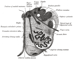
 Transverse section of medulla oblongata of human embryo. | |
| Details | |
| Identifiers | |
| Latin | Nucleus tractus solitarius medullae oblongatae. |
| MeSH | A08.186.211.132.810.406.750 |
| NeuroNames | hier-739 |
| NeuroLex ID | Solitary nucleus |
| TA | A14.1.04.230 |
| FMA | 72242 |
In the human brain, the solitary nucleus (nucleus of the solitary tract, nucleus solitarius, nucleus tractus solitarius, nucleus tractus solitarii, NTS) is a series of purely sensory nuclei (clusters of nerve cell bodies) forming a vertical column of grey matter embedded in the medulla oblongata. Through the center of the NTS runs the solitary tract, a white bundle of nerve fibers, including fibers from the facial, glossopharyngeal and vagus nerves, that innervate the NTS. The NTS projects to, among other regions, the reticular formation, parasympathetic preganglionic neurons, hypothalamus and thalamus, forming circuits that contribute to autonomic regulation. Cells along the length of the NTS are arranged roughly in accordance with function; for instance, cells involved in taste are located in the higher, more forward ("rostral") part, while those receiving information from cardio-respiratory and gastrointestinal processes are found in the lower, more posterior ("caudal") part.[1][2]
Inputs
- Taste information from the facial nerve (anterior 2/3 of the tongue), glossopharyngeal nerve (posterior 1/3) and vagus nerve (small area on the epiglottis)
- Sensory information from the ear (Auricular branch of the vagus nerve)
- Chemoreceptors and mechanoreceptors of the general visceral afferent pathway (GVA) in the carotid body via glossopharyngeal nerve, aortic bodies, and the atrial pacemaker, via the vagus nerve
- Chemically and mechanically sensitive neurons of the general visceral afferent pathway (GVA) with endings located in the heart, lungs, airways, gastrointestinal system, pharynx, and liver via the glossopharyngeal and vagus nerves
Neurons that innervate the NTS mediate the gag reflex, the carotid sinus reflex, the aortic reflex, the cough reflex, the baroreceptor and chemoreceptor reflexes, several respiratory reflexes and reflexes within the gastrointestinal system regulating motility and secretion.
Neurons which transmit signals about the gut wall, the stretch of the lungs, and the dryness of mucous membranes also innervate the NTS. The first central neurons within the NTS can participate in simple autonomic reflexes.
Outputs
Information goes from the NTS to a large number of other regions of the brain including the paraventricular nucleus of the hypothalamus and the central nucleus of the amygdala, as well as to other nuclei in the brainstem (such as the parabrachial area, the Locus coeruleus, the Dorsal raphe nucleus , and other visceral motor or respiratory networks).[3] The signals projected from the NTS to the parabrachial area originate in the oral cavity and gastrointestinal tract. The pathways for gastric and gustatory (taste) processes are believed to terminate in different subdivisions of the parabrachial area, but still interact in the NTS.[4][5] Some neuronal subpopulations in the NTS, such as the noradrenergic A2 neurons and the aldosterone-sensitive HSD2 neurons project as far rostrally as the bed nucleus of the stria terminalis.[6][7]
Additional images
 Section of the medulla oblongata at about the middle of the olive.
 Primary terminal nuclei of the afferent (sensory) cranial nerves schematically represented; lateral view.
See also
References
- ↑ Duane E. Haines (2004). Neuroanatomy: An Atlas of Structures, Sections, and Systems. Lippincott Williams & Wilkins. pp. 186–. ISBN 978-0-7817-4677-9. Retrieved 22 January 2013.
- ↑ P. Michael Conn (2008). Neuroscience in Medicine. Springer. p. 264. ISBN 978-1-60327-455-5. Retrieved 22 January 2013.
- ↑ Carlson, Neil R. (2010). Physiology of Behavior (10th ed.). Allyn & Bacon. p. 253. ISBN 978-0-205-66627-0.
- ↑ Karimnamazi, Hamid; Travers, Susan P; Travers, Joseph B (2002). "Oral and gastric input to the parabrachial nucleus of the rat". Brain Research. 957 (2): 193–206. doi:10.1016/S0006-8993(02)03438-8. PMID 12445962.
- ↑ Karimnamazi, Hamid; Travers, Joseph B. (1998). "Differential projections from gustatory responsive regions of the parabrachial nucleus to the medulla and forebrain". Brain Research. 813 (2): 283–302. doi:10.1016/S0006-8993(98)00951-2. PMID 9838165.
- ↑ Geerling JC, Loewy AD. Aldosterone-sensitive neurons in the nucleus of the solitary tract: efferent projections. J Comp Neurol. 2006 Jul 10;497(2):223-50. PMID 16705681
- ↑ Shin JW, Geerling JC, Loewy AD. Inputs to the ventrolateral bed nucleus of the stria terminalis. J Comp Neurol. 2008 Dec 10;511(5):628-57. doi: 10.1002/cne.21870. PMID 18853414