Pterygopalatine ganglion
| Pterygopalatine ganglion | |
|---|---|
 Alveolar branches of superior maxillary nerve and pterygopalatine ganglion | |
 The pterygopalatine ganglion and its branches (pterygopalatine ganglion visible but not labeled, as large yellow ganglion in upper-right center) | |
| Details | |
| From | Maxillary nerve and nerve of pterygoid canal |
| To | Greater palatine nerve, lesser palatine nerve, posterior lateral nasal branches and nasopalatine nerve |
| Identifiers | |
| Latin | Ganglion pterygopalatinum |
| TA | A14.3.02.006 |
| FMA | 6965 |
The pterygopalatine ganglion (Meckel's ganglion, nasal ganglion or sphenopalatine ganglion) is a parasympathetic ganglion found in the pterygopalatine fossa. It is largely innervated by the greater petrosal nerve (a branch of the facial nerve); and its axons project to the lacrimal glands and nasal mucosa. The flow of blood to the nasal mucosa, in particular the venous plexus of the conchae, is regulated by the pterygopalatine ganglion and heats or cools the air in the nose. It is one of four parasympathetic ganglia of the head and neck, the others being the submandibular ganglion, otic ganglion, and ciliary ganglion.
Structure
The pterygopalatine ganglion (of Meckel), the largest of the parasympathetic ganglia associated with the branches of the maxillary nerve, is deeply placed in the pterygopalatine fossa, close to the sphenopalatine foramen. It is triangular or heart-shaped, of a reddish-gray color, and is situated just below the maxillary nerve as it crosses the fossa.
The pterygopalatine ganglion supplies the lacrimal gland, paranasal sinuses, glands of the mucosa of the nasal cavity and pharynx, the gingiva, and the mucous membrane and glands of the hard palate. It communicates anteriorly with the nasopalatine nerve.
Roots
It receives a sensory, a parasympathetic, and a sympathetic root.
Sensory root
Its sensory root is derived from two sphenopalatine branches of the maxillary nerve; their fibers, for the most part, pass directly into the palatine nerves; a few, however, enter the ganglion, constituting its sensory root.
Parasympathetic root
Its parasympathetic root is derived from the nervus intermedius (a part of the facial nerve) through the greater petrosal nerve.
In the pterygopalatine ganglion, the preganglionic parasympathetic fibers from the greater petrosal branch of the facial nerve synapse with neurons whose postganglionic axons, vasodilator, and secretory fibers are distributed with the deep branches of the trigeminal nerve to the mucous membrane of the nose, soft palate, tonsils, uvula, roof of the mouth, upper lip and gums, and upper part of the pharynx. It also sends postganglionic parasympathetic fibers to the lacrimal nerve (a branch of the Ophthalmic nerve, also part of the trigeminal nerve) via the zygomatic nerve, a branch of the maxillary nerve (from the trigeminal nerve), which then arrives at the lacrimal gland.
The nasal glands are innervated with secretomotor from the greater petrosal nerve. Likewise, the palatine glands are innervated by the nasopalatine, greater palatine nerve and lesser palatine nerves. The pharyngeal nerve innervates pharyngeal glands. These are all branches of maxillary nerve.
Sympathetic root
The ganglion also consists of sympathetic efferent (postganglionic) fibers from the superior cervical ganglion. These fibers, from the superior cervical ganglion, travel through the carotid plexus, and then through the deep petrosal nerve. The deep petrosal nerve (carrying postganglionic sympathetics) joins with the greater petrosal nerve (carrying preganglionic parasympathetics) to form the nerve of the pterygoid canal, which passes through the pterygoid canal before entering the ganglion.
Branches
- Orbital branches? (See "Innervation" section of ethmoid sinus page.)
- Nasopalatine nerve
- Greater palatine nerve
- Lesser palatine nerve
- Medial and Lateral Posterior Superior and Posterior Inferior Nasal Branches
- Pharyngeal branch of maxillary nerve
Additional images
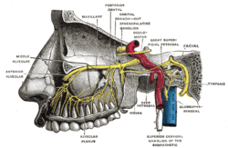
 Plan of the facial and intermediate nerves and their communication with other nerves.
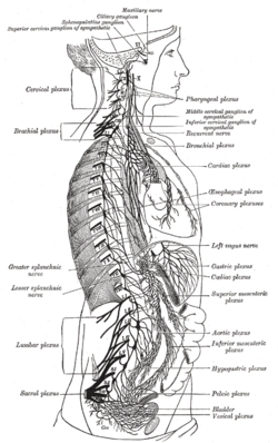 The right sympathetic chain and its connections with the thoracic, abdominal, and pelvic plexuses.
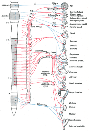 Diagram of efferent sympathetic nervous system.
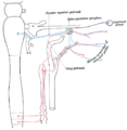 Sympathetic connections of the sphenopalatine and superior cervical ganglia.
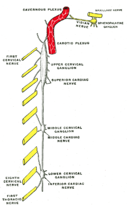 Diagram of the cervical sympathetic.
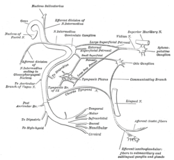
 An illustration of the path of the Maxillary nerve.
References
This article incorporates text in the public domain from the 20th edition of Gray's Anatomy (1918)
External links
- synd/2132 at Who Named It?
- cranialnerves at The Anatomy Lesson by Wesley Norman (Georgetown University) (V, VII)