Marginal mandibular branch of the facial nerve
| Marginal mandibular branch of the facial nerve | |
|---|---|
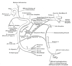 Plan of the facial and intermediate nerves and their communication with other nerves. (Labeled at center bottom, second from bottom, as "Mandibular".) | |
 The nerves of the scalp, face, and side of neck. | |
| Details | |
| From | facial nerve |
| Identifiers | |
| Latin | ramus marginalis mandibularis nervi facialis |
| TA | A14.2.01.113 |
| FMA | 53365 |
The marginal mandibular branch of the facial nerve passes forward beneath the platysma and depressor anguli oris, supplying the muscles of the lower lip and chin, and communicating with the mental branch of the inferior alveolar nerve.
Muscles innervated
The marginal mandibular branch innervates the following muscles:[1]
- Depressor labii inferioris - lowers bottom lip down and laterally. Origin: Anterior part of oblique line of mandible. Insertion: Lower lip at midline, blends with muscle from opposite side.
- Depressor anguli oris (triangularis) - lowers the corner of the mouth down and laterally. Origin: Oblique line of mandible below canine, premolar, and first molar teeth. Insertion: Skin at the corner of mouth and blending with orbicularis oris.
- Mentalis - raises and protrudes lower lip as it wrinkles skin on chin. Origin: Mandible inferior to incisor teeth. Insertion: Skin of chin.
Clinical significance
The marginal mandibular nerve may be injured during surgery in the neck region, especially during excision of the submandibular salivary gland or during neck dissections.
Additional images
 Lateral head anatomy detail
 Lateral head anatomy detail. Neonatal dissection.
References
This article incorporates text in the public domain from the 20th edition of Gray's Anatomy (1918)
External links
- Anatomy photo:23:06-0103 at the SUNY Downstate Medical Center - "Branches of Facial Nerve (CN VII)"
- lesson4 at The Anatomy Lesson by Wesley Norman (Georgetown University) (parotid3)
- cranialnerves at The Anatomy Lesson by Wesley Norman (Georgetown University) (VII)
- http://www.dartmouth.edu/~humananatomy/figures/chapter_47/47-5.HTM
This article is issued from Wikipedia - version of the 4/18/2015. The text is available under the Creative Commons Attribution/Share Alike but additional terms may apply for the media files.