Inferior cerebellar peduncle
| Inferior cerebellar peduncle | |
|---|---|
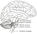 Scheme showing the connections of the several parts of the brain. (Inferior peduncle labeled at bottom right.) | |
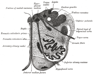 Section of the medulla oblongata at about the middle of the olive. (Inferior peduncle labeled at upper right. | |
| Details | |
| Identifiers | |
| Latin | pedunculus cerebellaris inferior |
| NeuroNames | hier-778 |
| NeuroLex ID | Inferior cerebellar peduncle |
| TA | A14.1.04.013 |
| FMA | 72615 |
The upper part of the posterior district of the medulla oblongata is occupied by the inferior cerebellar peduncle, a thick rope-like strand situated between the lower part of the fourth ventricle and the roots of the glossopharyngeal and vagus nerves.
Each cerebellar inferior peduncle connects the spinal cord and medulla oblongata with the cerebellum, and comprises the juxtarestiform body and restiform body.
Important fibers running through the inferior cerebellar peduncle include the dorsal spinocerebellar tract and axons from the inferior olivary nucleus, among others.
Function
The inferior cerebellar peduncle carries many types of input and output fibers that are mainly concerned with integrating proprioceptive sensory input with motor vestibular functions such as balance and posture maintenance. It consists of the following fiber tracts entering cerebellum:
- Posterior spinocerebellar tract: unconsciousness proprioceptive information from the lower part of trunk and lower limb. This tract originates at the ipsilateral Clarke's nucleus (T1-L1) and travels upward to reach the inferior cerebellar peduncle and synapses within the spinocerebellum (also known as the paleocerebellum).
- Cuneocerebellar tract: unconsciousness proprioceptive information from the upper limb and neck. This tract originates at the ipsilateral accessory cuneate nucleus and travels through the inferior cerebellar peduncle to reach the spinocerebellum part of the cerebellum.
- Trigeminocerebellar tract: unconsciousness proprioceptive information from the face.
- Olivocerebellar tract: "error signal" in movement originates from the cerebral cortex and spinal cord. This tract originates at contralateral inferior olivary nucleus and enters the cerebellum as a climbing fiber.
- Vestibulocerebellar tract: vestibular information projects onto the vestibulocerebellum (also known as the archicerebellum).
This peduncle also carries information leaving cerebellum: from the Purkinje cells to the vestibular nuclei in the dorsal brainstem located at the junction between the pons and medulla oblongata.
See also
Additional images
 Upper part of medulla spinalis and hind- and mid-brains; posterior aspect, exposed in situ.
 Superficial dissection of brain-stem. Lateral view.
 Dissection of brain-stem. Lateral view.
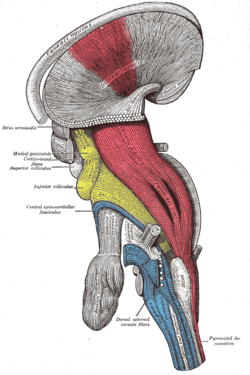 Deep dissection of brain-stem. Lateral view.
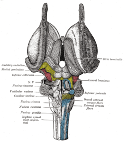 Dissection of brain-stem. Dorsal view.
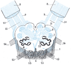 Diagram showing the course of the arcuate fibers.
 Dissection showing the course of the cerebrospinal fibers.
 Cross section of lower pons showing part of the inferior cerebellar peduncle (#8) labeled at the upper left.
 Cerebellum. Inferior surface.
 Cerebellum. Inferior surface.
 Cerebellum. Inferior surface.
References
This article incorporates text in the public domain from the 20th edition of Gray's Anatomy (1918)
External links
- Atlas image: n2a5p2 at the University of Michigan Health System
- Atlas image: n2a7p4 at the University of Michigan Health System
- Illustration and text: cere/text/p1/icp.htm at the University of Wisconsin-Madison Medical school