Submandibular duct
| Submandibular duct | |
|---|---|
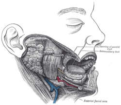 Dissection, showing salivary glands of right side. (Labeled as "submaxillary duct", but is identified as "submandibular duct" in newer sources.) | |
 Mandibular division of trifacial nerve, seen from the middle line. The small figure is an enlarged view of the otic ganglion. ("Wharton's duct" labeled in lower left.) | |
| Details | |
| Identifiers | |
| Latin | Ductus submaxillaris |
| MeSH | A03.556.500.760.640 |
| TA | A05.1.02.012 |
| FMA | 86266 |
The submandibular duct or Wharton duct[1] or submaxillary duct, is one of the salivary excretory ducts. It is about 5 cm. long, and its wall is much thinner than that of the parotid duct. It drains saliva from each bilateral submandibular gland and sublingual gland to the sublingual caruncle at the base of the tongue.[2]
Structure
It begins by numerous branches from the deep surface of the gland, and runs forward between the mylohyoid, hyoglossus, and genioglossus muscles. It then passes between the sublingual gland and the genioglossus and opens by a narrow opening on the summit of a small papilla at the side of the frenulum of the tongue.
It lies superior to lingual and hypoglossal nerves.
Function
This is the duct from which a hungry person, preparing to take a first bite of food, might accidentally eject a spray of salivary fluid, or, alternatively, intentionally do so in a process called gleeking.
History
It was initially described by the English anatomist Thomas Wharton and is sometimes referred to by his name.[3]
References
This article incorporates text in the public domain from the 20th edition of Gray's Anatomy (1918)
- ↑ Wharton's duct at Who Named It?
- ↑ Ten Cate's Oral Histology, Nanci, Elsevier, 2013, page 255
- ↑ Wharton T (1656). Adenographia: sive glandularum totius corporis descriptio. London: Wharton. pp. 128–137.
External links
- Anatomy figure: 34:03-05 at Human Anatomy Online, SUNY Downstate Medical Center
- MedicalMnemonics.com: 329