Iliacus muscle
| Iliacus muscle | |
|---|---|
 Position of iliacus muscle (shown in red.) | |
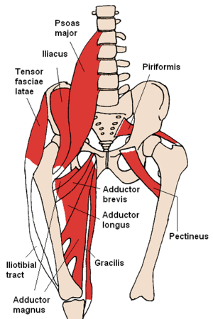 The iliacus and nearby muscles | |
| Details | |
| Origin | upper two-third of the iliac fossa |
| Insertion | base of the lesser trochanter of femur |
| Artery | medial femoral circumflex artery, iliac branch of iliolumbar artery |
| Nerve | femoral nerve |
| Actions | flexes and rotates laterally thigh |
| Antagonist | Gluteus maximus |
| Identifiers | |
| Latin | musculus iliacus |
| TA | A04.7.02.003 |
| FMA | 22310 |
The iliacus (/ᵻˈlaɪ.əkəs/) is a flat, triangular muscle which fills the iliac fossa.
Structure
The iliacus arises from the iliac fossa on the interior side of the hip bone, and also from the region of the anterior inferior iliac spine (AIIS). It joins the psoas major to form the Iliopsoas as which it proceeds across the iliopubic eminence through the muscular lacuna to its insertion on the lesser trochanter of the femur. Its fibers are often inserted in front of those of the psoas major and extends distally over the lesser trochanter. [1]
Innervation
The iliopsoas is innervated by the femoral nerve and direct branches from the lumbar plexus.[2]
Function
In open-chain exercises, as part of the iliopsoas, the iliacus is important for lifting (flexing) the femur forward (i.e. front scale). In closed-chain exercises, the iliopsoas bends the trunk forward and can lift the trunk from a lying posture (i.e. sit-ups, back scale) because the psoas major crosses several vertebral joints and the sacroiliac joint. From its origin in the lesser pelvis the iliacus acts exclusively on the hip joint.[1]
Additional images
 Position of iliacus muscle (shown in red.) Animation.
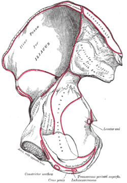 Right hip bone. Internal surface. (Iliac fossa visible at upper left.)
Notes
References
- Platzer, Werner (2004). Color Atlas of Human Anatomy, Vol. 1: Locomotor System (5th ed.). Thieme. ISBN 3-13-533305-1.
- Thieme Atlas of Anatomy: General Anatomy and Musculoskeletal System. Thieme. 2006. ISBN 1-58890-419-9.
External links
| Wikimedia Commons has media related to Iliacus muscles. |
- -181075889 at GPnotebook
- PTCentral
- Anatomy figure: 40:07-05 at Human Anatomy Online, SUNY Downstate Medical Center - "Muscles and nerves of the posterior abdominal wall."
- pelvis at The Anatomy Lesson by Wesley Norman (Georgetown University) (femalepelvicdiaphragm, malepelvicdiaphragm)