Anatomical terms of muscle
 |
| This article is part of a series on |
| Anatomical terminology |
|---|
| Motion |
| Location |
| Muscle |
| Bone |
| Neuroanatomy |
Muscles are described using unique anatomical terminology according to their actions and structure.
Classification
There are three types of muscle tissue in the human body: skeletal, smooth and cardiac.
Skeletal muscle
Skeletal striated muscle, or Voluntary muscle, primarily links to bone through tendon, thus enabling through levers of the bones of the human skeleton posturing, upright stance under atmospheric pressure, and voluntary movements.[1]
Smooth muscle
Smooth muscle tissue is found in parts of the body where it conveys action without conscious intent. The majority of this type of muscle tissue is found in the Digestive and Urinary systems where it acts by propelling forward food, chyme, and feces in the former and urine in the latter. Other places smooth muscle can be found are within the uterus, where it helps facilitate birth, and the eye, where the pupillary sphincter controls pupil size.[2]
Cardiac muscle
Cardiac muscle is specific to the heart. It is also involuntary in its movement, and is additionally self-excitatory, contracting without outside stimuli.[3]
Actions of skeletal muscle
.jpg)
As well as anatomical terms of motion, which describe the motion made by a muscle, unique terminology is used to describe the action of a set of muscles.
Agonists and antagonists
Agonist muscles and antagonist muscles refer to muscles that cause or inhibit a movement.
Agonist muscles cause a movement to occur through their own contraction. [4] For example, the triceps brachii contracts during the up phase of a push-up (elbow extension). During the down phase of a push-up, the same triceps brachii actively controls elbow flexion while relaxing. It is still the agonist, because while resisting gravity during relaxing, the triceps brachii continues to be the prime mover, or controller, of the joint action. (Agonists are also interchangeably referred to as "prime movers," since they are the muscles considered primarily responsible for generating a specific movement. This term typically describes skeletal muscles.[5])
Antagonist muscles oppose a specific movement. [6] This controls a motion, slows it down, and returns a limb to its initial position. Antagonism is not an intrinsic property; it is a role that a muscle plays depending on the motion. If a motion is reversed, agonist and antagonist muscles switch roles. Because a flexor muscle is always a flexor, in flexion it is the agonist, and in extension it is the antagonist. Conversely, an extensor muscle is the agonist in extension and the antagonist in flexion. Using the example above of the triceps brachii during a push-up, the elbow flexor muscles are the antagonists during both the up phase and down phase of the movement.
Agonist-antagonist pairs
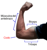
Antagonist and agonist muscles often occur in pairs, called antagonistic pairs. As one muscle contracts, the other relaxes. An example of an antagonisic pair is the biceps and triceps; to contract - the triceps relaxes while the biceps contracts to lift the arm. "Reverse motions" need antagonistic pairs located in opposite sides of a joint or bone, including abductor-adductor pairs and flexor-extensor pairs. These consist of an extensor muscle, which "opens" the joint (by increasing the angle between the two bones) and a flexor muscle, which does the opposite by decreasing the angle between two bones.
However muscles don't always work this way - sometimes agonists and antagonists contract at the same time to produce force, as per Lombard's paradox.
Not all muscles are paired in this way. An example of exception is the deltoid.
Synergistic action
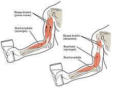
Synergist muscles perform, or help perform, the same set of joint motion as the agonists. Synergists muscles act on movable joints. Synergists are sometimes referred to as "neutralizers" because they help cancel out, or neutralize, extra motion from the agonists to make sure that the force generated works within the desired plane of motion.
Muscle fibers can only contract up to 40% of their fully stretched length. Thus the short fibers of pennate muscles are more suitable where power rather than range of contraction is required. This limitation in the range of contraction affects all muscles, and those that act over several joints may be unable to shorten sufficiently to produce the full range of movement at all of them simultaneously (active insufficiency, e.g., the fingers cannot be fully flexed when the wrist is also flexed). Likewise, the opposing muscles may be unable to stretch sufficiently to allow such movement to take place (passive insufficiency). For both these reasons, it is often essential to use other muscles, called fixators or synergists, in this type of action to fix certain of the joints so that others can be moved effectively, e.g., fixation of the wrist during full flexion of the fingers in clenching the fist. Synergists are muscles that facilitate the fixation action.
There is an important difference between a helping synergist muscle and a true synergist muscle. A true synergist muscle is one that only neutralizes an undesired joint action, whereas a helping synergist is one that neutralizes an undesired action but also assists with the desired action.
Neutralizer Action
A muscle that fixes or holds a bone so that the agonist can carry out the intended movement is said to have a neutralising action. A good famous example of this are the hamstrings; the semitendinosus and semimembranosus muscles perform knee flexion and knee internal rotation whereas the biceps femoris carries out knee flexion and knee external rotation. For the knee to flex while not rotating in either direction, all three muscles contract to stabilize the knee while it moves in the desired way.
Composite muscle
Composite or hybrid muscles have more than one set of fibers that perform the same function, and are usually supplied by different nerves for different set of fibers. For example, the tongue itself is a composite muscle made up of various components like longitudinal, transverse, horizontal muscles with different parts innervated having different nerve supply.
Actions of smooth muscle
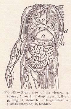
Smooth muscles are responsible for the contract-ability of hollow organs, such as blood vessels, the gastrointestinal tract, the bladder, or the uterus. It can develop isometric force per cross-sectional area that is equal to that of skeletal muscle. However, the speed of smooth muscle contraction is only a small fraction of that of skeletal muscle.[7]
Ca2+ Dependent contraction of smooth muscle
Contraction of smooth muscle is initiated by a Ca2+.
Smooth muscle relaxation
Smooth muscle relaxation occurs either as a result of removal of the contractile stimulus or by the direct action of a substance that stimulates inhibition of the contractile mechanism (e.g., atrial natriuretic factor is a vasodilator).
Form
Insertion and origin
The insertion and origin of a muscle are the two places where it is anchored, one at each end. The tissue of the attachment is called an enthesis.
The origin of a muscle is the bone, typically proximal, which has greater mass and is more stable during a contraction than a muscle's insertion. [8] For example, with the latissimus dorsi muscle, the origin site is the torso, and the insertion is the arm. When this muscle contracts, normally the arm moves due to having less mass than the torso. This is the case when grabbing objects lighter than the body, as in the typical use of a lat pull down machine. This can be reversed however, such as in a chin up where the torso moves up to meet the arm.
The insertion of a muscle is the structure that it attaches to and tends to be moved by the contraction of the muscle. [9] This may be a bone, a tendon or the subcutaneous dermal connective tissue. Insertions are usually connections of muscle via tendon to bone.[10] The insertion is a bone that tends to be distal, have less mass, and greater motion than the origin during a contraction.
Muscle fibres
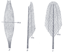
A: fusiform. B: unipennate. C: bipennate.
(P.C.S., physiological cross-section)
Muscles may also be described by the direction that the muscle fibres run in.
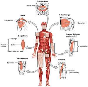
- Fusiform muscles have fibres that run parallel to the length of the muscle, and are spindle-shaped. [11] For example, the pronator teres muscle of the forearm.
- Unipennate muscles have fibres that run the entire length of only one side of a muscle, like a quill pen. For example, the fibularis muscles.
- Bipennate muscles consist of two rows of oblique muscle fibres, facing in opposite diagonal directions, converging on a central tendon. Bipennate muscle is stronger than both unipennate muscle and fusiform muscle, due to a larger physiological cross-sectional area. Bipennate muscle shortens less than unipennate muscle but develops greater tension when it does, translated into greater power but less range of motion. Pennate muscles generally also tire easily. Examples of bipennate muscles are the rectus femoris muscle of the thigh, and the stapedius muscle of the middle ear.
State
Hypertrophy and atrophy
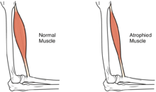
Hypertrophy is increase in muscle size from an increase in size of individual muscle cells. This usually occurs as a result of exercise.
See also
- Reciprocal inhibition
- Anatomical terms of location
- Anatomical terms of motion
- Anatomical terms of bone
- Anatomical terms of neuroanatomy
References
This article incorporates text in the public domain from the 20th edition of Gray's Anatomy (1918)
- ↑ Skeletal Muscle
- ↑ Smooth Muscle
- ↑ Cardiac Muscle
- ↑ Taber 2001, pp. "Agonist".
- ↑ Baechle, Thomas (2008). Essentials of Strength Training and Conditioning. USA: National Strength and Conditioning Association. ISBN 978-0-7360-8465-9.
- ↑ Taber 2001, pp. "Antagonist".
- ↑ SMOOTH MUSCLE - The university Of Illinois At Chicago Archived November 18, 2015, at the Wayback Machine.
- ↑ OED 1989, "origin".
- ↑ Taber 2001, "insertion".
- ↑ Martini, Frederic; William C. Ober; Claire W. Garrison; Kathleen Welch; Ralph T. Hutchings (2001). Fundamentals of Anatomy and Physiology, 5th Ed. Prentice Hall. ISBN 0130172928.
- ↑ Taber 2001, "Fusiform".
- Books
- Willert, editor Donald Venes, Coeditor Clayton L. Thomas, Managing Editor Elizabeth J. Egan, Assistant Editors Nancee A. Morelli and Alison D. Nell. /copy Editor Ann Houska Proofreaders Joy Matkowski and Christopher Muldor. Dictionary Illustrator Beth Anne (2001). Taber's cyclopedic medical dictionary. (Ed. 19, illustrated in full color ed.). Philadelphia: F.A.Davis Co. ISBN 0-8036-0655-9.
- J. A. Simpson, ed. (1989). The Oxford English dictionary. Oxford: Clarendon Press. ISBN 9780198611868.