Hamstring
| Hams | |
|---|---|
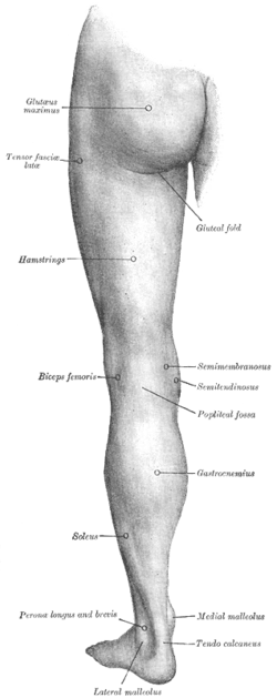 Posterior view of left lower extremity. | |
| Details | |
| Origin | tuberosity of the ischium, linea aspera |
| Insertion | tibia, fibula |
| Artery | inferior gluteal artery, profunda femoris artery |
| Nerve | sciatic nerve (tibial nerve and common fibular nerve)[1][2] |
| Actions | flexion of knee, extension of hip |
| Antagonist | Rectus femoris muscle |
In human anatomy, a hamstring is one of the three posterior thigh muscles (from medial to lateral: semitendinosus, semimembranosus and biceps femoris).[3]
In quadrupeds, the hamstring is the single large tendon found behind the knee or comparable area.
Criteria
The common criteria of any hamstring muscles are:
- Muscles should originate from ischial tuberosity.
- Muscles should be inserted over the knee joint, in the tibia or in the fibula.
- Muscles will be innervated by the tibial branch of the Sciatic nerve.
- Muscle will participate in flexion of the knee joint and extension of the hip joint.
Those muscle which fulfills all of the four criteria are called true hamstrings.
The adductor magnus reaches only up to the adductor tubercle of the femur, but it is included amongst the hamstrings because the tibial collateral ligament of the knee joint morphologically is the degenerated tendon of this muscle. The ligament is attached to medial epicondyle, two millimeters from the adductor tubercle.
Structure
The three muscles of the posterior thigh (semitendinosus, semimembranosus, biceps femoris long & short head) flex (bend) the knee, while all but the short head of biceps femoris extend (straighten) the hip. The three 'true' hamstrings cross both the hip and the knee joint and are therefore involved in knee flexion and hip extension. The short head of the biceps femoris crosses only one joint (knee) and is therefore not involved in hip extension. With its divergent origin and innervation it is sometimes excluded from the 'hamstring' characterization.[4]
| Muscle | Origin | Insertion | Nerve |
| semitendinosus | ischial tuberosity | medial surface of tibia | tibial part of sciatic |
| semimembranosus | ischial tuberosity | medial tibial condyle | tibial part of sciatic |
| biceps femoris - long head | ischial tuberosity | lateral side of the head of the fibula | tibial part of sciatic |
| biceps femoris - short head | linea aspera and lateral supracondylar line of femur | lateral side of the head of the fibula (common tendon with the long head) | common peroneal |
A portion of the adductor magnus is sometimes considered a part of the hamstrings.[4]
Function
The hamstrings cross and act upon two joints - the hip and the knee.
Semitendinosus and semimembranosus extend the hip when the trunk is fixed; they also flex the knee and medially (inwardly) rotate the lower leg when the knee is bent.
The long head of the biceps femoris extends the hip, as when beginning to walk; both short and long heads flex the knee and laterally (outwardly) rotate the lower leg when the knee is bent.
The hamstrings play a crucial role in many daily activities such as walking, running, jumping, and controlling some movement in the trunk. In walking, they are most important as an antagonist to the quadriceps in the deceleration of knee extension.
Clinical significance
Imaging

Imaging the hamstring muscles is usually performed with an ultrasound and/or MRI.[5] The biceps femoris is most commonly injured, followed by semitendinosus. Semimembranosus injury is rare. Imaging is useful in differentiating the grade of strain, especially if the muscle is completely torn.[6] In this setting, the level and degree of retraction can be determined, serving as a useful roadmap prior to any surgery. Those with a hamstring strain of greater than 60mm in length have a greater risk of recurrence.[7]
Use in surgery
The distal semitendinosus tendon is one of the tendons that can be used in the surgical procedure ACL reconstruction. In this procedure, a piece of it is used to replace the anterior cruciate ligament (ACL). The ACL is one of the four major ligaments in the knee.
History
Etymology
The word "ham" is derived from the Old English ham or hom meaning the hollow or bend of the knee, from a Germanic base where it meant "crooked". It gained the meaning of the leg of an animal around the 15th century.[8] String refers to tendons, and thus, the hamstrings are the string-like tendons felt on either side of the back of the knee.[9]
See also
References
- ↑ University of Glasgow :: Biomedical & Life Sciences :: Biomedical & Life Sciences
- ↑ "Biceps Femoris - Short Head — Musculoskeletal Radiology — UW Radiology". Rad.washington.edu. Retrieved 2012-11-02.
- ↑ Mayo Clinic Staff (3 Oct 2015). "Hamstring injury". Mayo clinic. Retrieved 6 July 2016.
- 1 2 postthigh at The Anatomy Lesson by Wesley Norman (Georgetown University)
- ↑ Koulouris G, Connell D (2003). "Evaluation of the hamstring muscle complex following acute injury". Skeletal Radiol. 32 (10): 582–9. doi:10.1007/s00256-003-0674-5. PMID 12942206.
- ↑ Schache AG, Koulouris G, Kofoed W, Morris HG, Pandy MG (2008). "Rupture of the conjoint tendon at the proximal musculotendinous junction of the biceps femoris long head: a case report.". Knee Surg Sports Traumatol Arthrosc. 16 (8): 797–802. doi:10.1007/s00167-008-0517-y. PMID 18360748.
- ↑ Koulouris G, Connell DA, Brukner P, Schneider-Kolsky M (2007). "Magnetic resonance imaging parameters for assessing risk of recurrent hamstring injuries in elite athletes.". Am J Sports Med. 35 (9): 1500–6. doi:10.1177/0363546507301258. PMID 17426283.
- ↑ Brown, Lesley, ed. (2007). Shorter Oxford English Dictionary II (Sixth ed.). Oxford: Oxford University press. p. 3611.
- ↑ "Online Etymology Dictionary". Etymonline.com. Retrieved 2012-11-02.