Gastrocnemius muscle
| Gastrocnemius muscle | |
|---|---|
 | |
| Details | |
| Origin | superior to articular surfaces of lateral condyle of femur and medial condyle of femur |
| Insertion | tendo calcaneus (achilles tendon) into mid-posterior calcaneus |
| Artery | sural arteries |
| Nerve | tibial nerve from the sciatic, specifically, nerve roots S1–S2 |
| Actions | plantar flexes foot, flexes knee |
| Antagonist | Tibialis anterior muscle |
| Identifiers | |
| TA | A04.7.02.044 |
| FMA | 22541 |
In humans, the gastrocnemius muscle (/ˌɡæstrɒkˈniːmiəs/ or /ˌɡæstrəkˈniːmiəs/; plural gastrocnemii; Latin, from Greek γαστήρ "stomach" and κνήμη (knēmē) "leg"; meaning "stomach of leg" (referring to the bulging shape of the calf) is a very powerful superficial bipennate muscle that is in the back part of the lower leg. It runs from its two heads just above the knee to the heel, a two joint muscle.
Structure
The gastrocnemius is located with the soleus in the posterior (back) compartment of the leg. The lateral head originates from the lateral condyle of the femur, while the medial head originates from the medial condyle of the femur. Its other end forms a common tendon with the soleus muscle; this tendon is known as the calcaneal tendon or Achilles Tendon and inserts onto the posterior surface of the calcaneus, or heel bone.
Deep to the gastrocnemius (farther from the skin) is the soleus muscle. Some anatomists consider both to be a single muscle, the triceps surae or "three-headed [muscle] of the calf", since they share a common insertion via the Achilles tendon. The plantaris muscle and a portion of its tendon run between the two muscles, which is involved in "locking" the knee from the standing position. Since the anterior compartment of the leg is lateral to the tibia, the bulge of muscle medial to the tibia on the anterior side is actually the posterior compartment. The soleus is superficial to the mid-shaft of the tibia.
Variation
10% to 30% of individuals have a sesamoid bone called the "fabella" in the lateral (outer) head of gastrocnemius muscle.
Function
The gastrocnemius is primarily involved in running, jumping and other "fast" movements of leg, and to a lesser degree in walking and standing. This specialization is connected to the predominance of white muscle fibers (type II fast twitch) present in the gastrocnemius, as opposed to the soleus, which has more red muscle fibers (type I slow twitch) and is the primary active muscle when standing still, as determined by EMG studies.[1][2]
Along with the soleus muscle, it forms half of the calf muscle. Its function is plantar flexing the foot at the ankle joint and flexing the leg at the knee joint.
Motor pathway
The plan to use the gastrocnemius in running, jumping, knee and plantar flexing is created in the precentral gyrus in the cerebrum of the brain.[3] Once a plan is produced, the signal is sent to and down an upper motor neuron. The signal is passed through the internal capsule and decussates, or crosses, in the medulla oblongata, specifically in the lateral corticospinal track.[4] The signal continues down through the anterior horn of the spinal cord where the upper motor neuron synapses with the lower motor neuron. Signal propagation continues down the anterior rami (Lumbar 4-5 and Sacral 1-5) of the sacral plexus. The sciatic nerve branches off of the sacral plexus in which the tibial and common fibular nerves are wrapped in one sheath. The tibial nerve eventually separates from the sciatic nerve and innervates the gastrocnemius muscle. Thus, completing the plan the brain had originally started with, so that the actions of running, standing, and jumping could be executed.
Clinical significance
The gastrocnemius muscle is prone to spasms, which are painful, involuntary, contractions of the muscle that may last several minutes.[5]
A severe ankle dorsiflexion force may result in an injury of the muscle, commonly referred to as a "torn" or "strained" calf muscle, which is acutely painful and disabling.

The gastrocnemius muscle may also become inflamed due to overuse. Anti-inflammatory medications and physical therapy (heat, massage, and stretching) may be useful.
Anatomical abnormalities involving the medial head of gastrocnemius muscle result in popliteal artery entrapment syndrome.
History
In a 1967 EMG study, Herman and Bragin concluded that its most important role was plantar flexing in large contractions and in rapid development of tension.[2]
Additional images
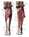 Nerves, arteries and veins surround the gastrocnemius and soleus.
 Muscle layer under the gastrocnemius
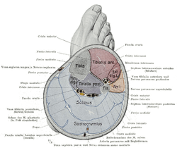 Cross section of the lower leg
 Right femur. Posterior surface.
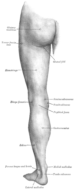 Back of left lower extremity.
 Gastrocnemius muscle
 Gastrocnemius muscle
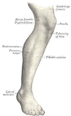 Lateral aspect of right leg.
References
- ↑ Clinically Oriented Anatomy. pp. 598–600. ISBN 978-1-60547-652-0.
- 1 2 Hamilton, Nancy; Luttgens, Kathryn (2001). Kinesiology: Scientific Basis of Human Motion (10th ed.). McGraw-Hill. ISBN 978-0-07-248910-1.
- ↑ Freberg, Laura (2015-01-01). Discovering Behavioral Neuroscience: An Introduction to Biological Psychology. Cengage Learning. ISBN 9781305687738.
- ↑ Saladin, Kenneth (2015). Anatomy & Physiology: The Unity of Form and Function. McGraw-Hill Education.
- ↑ "Nighttime Leg Cramps". WebMD. August 19, 2010. Retrieved March 7, 2012.
External links
| Wikimedia Commons has media related to Gastrocnemius muscle. |
- -275120049 at GPnotebook
- Anatomy photo:14:st-0405 at the SUNY Downstate Medical Center