Canine space
| Canine space | |
|---|---|
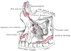 Lateral view of the maxilla, showing infra-orbital area, including the canine fossa, and muscle attachments. | |
The canine space (also termed the infra-orbital space),[1] is a fascial space of the head and neck (sometimes also termed fascial spaces or tissue spaces). It is a thin potential space on the face, and is paired on either side. It is located between the levator anguli oris muscle inferiorly and the levator labii superioris muscle superiorly.[1][2] The term is derived from the fact that the space is in the region of the canine fossa, and that infections originating from the maxillary canine tooth may spread to involve the space. Infra-orbital is derived from infra- meaning below and orbit which refers to the eye socket.
.png)
.png)
Structure
Boundaries
The boundaries of the canine space are:[2]
- the nasal cartilages anteriorly
- the buccal space posteriorly
- the quadratus labii superioris muscle (levator labii superioris) superiorly
- the oral mucosa of the maxillary labial sulcus inferiorly
- the quadratus labii superioris muscle superficially
- and the deep border is created by the levator anguli oris muscle.
Communications
the canine space communicates with the buccal space posteriorly.[2]
Function
Contents
The contents of the canine space are:[2]
- the angular artery and angular vein
- the infra-orbital nerve (a branch of the maxillary division of the trigeminal nerve)
Clinical significance
Canine space infections may occur by spread of infection from the buccal space.[2] Signs and symptoms of a canine space abscess might include swelling that obliterates the nasolabial fold. If left untreated, infections of this space will eventually spontaneously drain via the medial or lateral canthus of the eye, as this is the path of least resistance.[2] Treatment is usually by surgical incision and drainage, and the incision is placed inside the mouth to avoid a facial scar.
Rarely, when infections of the canine space erode into the infra-orbital vein or the inferior ophthalmic vein (via the sinuses), there can be spread via the common ophthalmic vein through the superior orbital fissure and into the cavernous sinus. This can result in septic cavernous sinus thrombosis, which is a rare, but life-threatening condition.[2]
Odontogenic infection
Odontogenic infections may spread to involve the canine space. The most likely causative tooth is the maxillary canine or maxillary first premolar.[1] This occurs when pus (e.g. from a periapical abscess), perforates the buccal cortical plate of the maxilla above the level of attachment of the levator anguli oris muscle. This is more likely if the tooth root is long (the maxillary canine has the longest root of all the teeth), and its apex lies at a level above the muscle attachment.[1]
References
- 1 2 3 4 Hargreaves KM; Cohen S, eds. (2010). Cohen's pathways of the pulp. Berman LH (web editor) (10th ed.). St. Louis, Mo.: Mosby Elsevier. pp. 590–594. ISBN 978-0-323-06489-7.
- 1 2 3 4 5 6 7 Hupp JR, Ellis E, Tucker MR (2008). Contemporary oral and maxillofacial surgery (5th ed.). St. Louis, Mo.: Mosby Elsevier. pp. 317–333. ISBN 9780323049030.