Popliteal fossa
| Popliteal fossa | |
|---|---|
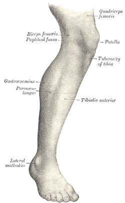 Lateral aspect of right leg | |
 Lymph glands of popliteal fossa | |
| Details | |
| Latin | Fossa poplitea |
The popliteal fossa (sometimes referred to as the kneepit) is a shallow depression located at the back of the knee joint. The bones of the popliteal fossa are the femur and the tibia.
Boundaries
The boundaries of the fossa are:[1]
| Medial | Lateral | |
|---|---|---|
| Superior | superior and medial: the semimembranosus muscle | superior and lateral: the biceps femoris muscle |
| Inferior | inferior and medial: the medial head of the gastrocnemius muscle | inferior and lateral: the lateral head of the gastrocnemius muscle and plantaris muscle |
Roof
The roof is formed by (from superficial to deep):[1]
- skin
- superficial fascia, which contains the small saphenous vein, the terminal branch of the posterior cutaneous nerve of the thigh, posterior division of the medial cutaneous nerve, lateral sural cutaneous nerve, and medial sural cutaneous nerve
- deep fascia or popliteal fascia
Floor
The floor is formed by:[1]
- the popliteal surface of the femur
- the capsule of the knee joint and the oblique popliteal ligament
- strong fascia covering the popliteus muscle
Contents
Structures within the popliteal fossa include, (from superficial to deep):[1]
- tibial nerve
- popliteal vein
- popliteal artery, a continuation of the femoral artery
- small saphenous vein (termination)[2]
- common fibular nerve (also known as the common peroneal nerve)[2]
- Popliteal lymph nodes and Dharmy vessels[2]
It is of note that the common fibular nerve also begins at the superior angle of the popliteal fossa.[3]
Additional images
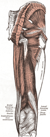 Muscles of the gluteal and posterior femoral regions.
 Small saphenous vein and its tributaries.
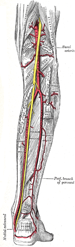 The popliteal, posterior tibial, and peroneal arteries.
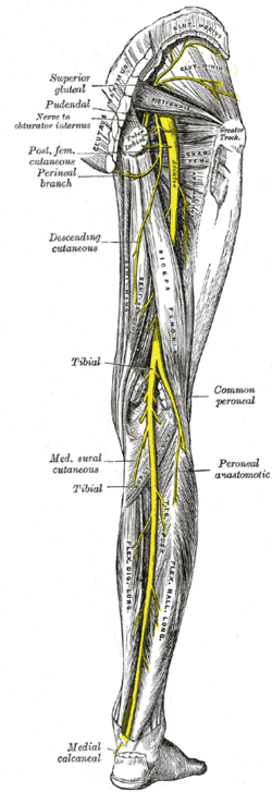 Nerves of the right lower extremity Posterior view.
 Muscles of thigh. Lateral view.
See also
References
- 1 2 3 4 Buckenmaier III C; Bleckner L (2008). "Chapter 20: Popliteal nerve block" (PDF). The Military Advanced Regional Anesthesia and Analgesia Handbook. Rockville, Maryland: Defense & Veterans Pain Management Initiative (DVPMI). Retrieved 2011-06-08.
- 1 2 3 Clinically Oriented Anatomy by Moore, 6th edition
- ↑ http://teachmeanatomy.info/lower-limb/areas/popliteal-fossa/
External links
- postthigh at The Anatomy Lesson by Wesley Norman (Georgetown University) (poplitealfossabones, poplitealfossacontents, poplitealfossafloor)
- MedicalMnemonics.com: 2747 9
This article is issued from Wikipedia - version of the 1/26/2016. The text is available under the Creative Commons Attribution/Share Alike but additional terms may apply for the media files.