Live cell imaging

Live cell imaging is the study of living cells using time-lapse microscopy. It is used by scientists to obtain a better understanding of biological function through the study of cellular dynamics.[1] Live cell imaging was pioneered in first decade of the 20th century. One of the first time-lapse microcinematographic films of cells ever made was made by Julius Ries, showing the fertilization and development of the sea urchin egg.[2] Since then, several microscopy methods have been developed which allow researchers to study living cells in greater detail with less effort. A newer type of imaging utilizing quantum dots have been used as they are shown to be more stable.
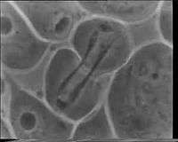


Phase contrast microscopy
Before the introduction of the phase contrast microscope it was difficult to observe living cells. As living cells are translucent they must be stained to be visible in a traditional light microscope. Unfortunately, the process of staining cells generally kill the cells. With the invention of the phase contrast microscopy it became possible to observe unstained living cells in detail. After its introduction in the 1940s, live cell imaging rapidly became popular using phase contrast microscopy.[6] The phase contrast microscope was popularized through a series of time-lapse movies (Video 1), recorded using a photographic film camera.[7] Its inventor, Frits Zernike, was awarded the Nobel Prize in 1953.[8] Other later phase contrast techniques used to observe unstained cells are Hoffman modulation and differential interference contrast microscopy.
Fluorescent microscopy
Phase contrast microscopy does not have the capacity to observe specific proteins or other organic chemical compounds which form the complex machinery of a cell. Synthetic and organic fluorescent stains have therefore been developed to label such compounds, making them observable by fluorescent microscopy (Video 2). Fluorescent stains are, however, phototoxic, invasive and bleach when observed. This limits their use when observing living cells over extended periods of time. Non-invasive phase contrast techniques are therefore often used as a vital complement to fluorescent microscopy in live cell imaging applications.[9][10]
Quantitative phase contrast microscopy
As a result of the rapid increase in pixel density of digital image sensors, quantitative phase contrast microscopy has emerged as an alternative microscopy method for live cell imaging.[11][12] Quantitative phase contrast microscopy has an advantage over fluorescent and phase contrast microscopy in that it is both non-invasive and quantitative in its nature. Contrary to phase contrast images, quantitative phase contrast images (Video 3) can be automatically processed to extract vast amount of dynamic cellular data from time-lapse image sequences.[13][14]
Due to the narrow focal depth of conventional microscopy, live cell imaging is to a large extent currently limited to observing cells on a single plane. Most implementations of quantitative phase contrast microscopy allow for images to be created and focused at different focal planes from a single exposure. This opens up the future possibility of 3-dimensional live cell imaging by means of fluorescence techniques.[15] A marker-free holographic technique that instead relies on complex deconvolution has been implemented and commercialized, which provides access to the non-invasive 3-dimensional imaging of live single cells.[16]
See also
References
- ↑ Monya Baker (2010). "Cellular imaging: Taking a long, hard look". Nature. 466 (26): 1137–1140. doi:10.1038/4661137a. PMID 20740018.
- ↑ Hannah Landecker (2009). "Seeing things: from microcinematography to live cell imaging". Nature Methods. 6 (10): 707–709. doi:10.1038/nmeth1009-707. PMID 19953685.
- ↑ Kurt Michel. "Historic phase contrast microscopy movies". The Cell — an image library. Archived from the original on October 6, 2014.
- ↑ George von Dassow; Koen J.C. Verbrugghe; Ann L. Miller; Jenny R. Sider; William M. Bement. "Cellular division in purple urchin embryo". The Cell — an image library.
- ↑ Birgit Janicke. "Digital holographic microscopy video showing cell division of unlabeled JIMT-1 breast cancer cells". The Cell — an image library.
- ↑ Mark Burgess (15 October 2003). "Celebrating 50 years of Live Cell Imaging" (PDF). Carl Zeiss UK and The Royal Microscopical Society. London: The Biochemical Society.
- ↑ Heinz Gundlach. "50 Years Ago: Frits Zernike (1888-1966) Got the Nobel Price in Physics for the Development of the Phase Contrast Method" (PDF) (Press release). Carl Zeiss AG. Archived from the original (PDF) on March 22, 2014.
- ↑ "The Nobel Prize in Physics 1953". Nobel Media AB.
- ↑ David J. Stephens; Victoria J. Allan (2003). "Light Microscopy Techniques for Live Cell Imaging". Science. 300 (5616): 82–86. doi:10.1126/science.1082160. PMID 12677057.
- ↑ Jing Ge; David K. Wood; David M. Weingeist; Somsak Prasongtanakij; Panida Navasumrit; Mathuros Ruchirawat; Bevin P. Engelward (2013). "Standard fluorescent imaging of live cells is highly genotoxic". Cytometry Part A. 83A (6): 552–560. doi:10.1002/cyto.a.22291.
- ↑ Etienne Cuche; Frédéric Bevilacqua; Christian Depeursinge (1999). "Digital holography for quantitative phase-contrast imaging". Optics Letters. 24 (5): 291–293. doi:10.1364/OL.24.000291.
- ↑ Pierre Marquet; Benjamin Rappaz; Pierre J. Magistretti; Etienne Cuche; Yves Emery; Tristan Colomb; Christian Depeursinge (2005). "Digital holographic microscopy: a noninvasive contrast imaging technique allowing quantitative visualization of living cells with subwavelength axial accuracy". Optics Letters. 30 (5): 468–470. doi:10.1364/OL.30.000468.
- ↑ "Label-free live cell time-lapse imaging & analysis". Phase Holographic Imaging AB. Archived from the original on April 20, 2013.
- ↑ "Quantitative phase contrast microscopy". Phase Holographic Imaging AB.
- ↑ Joseph Rosen; Gary Brooker (2008). "Non-scanning motionless fluorescence three-dimensional holographic microscopy". Nature Photonics. 2 (3): 190–195. doi:10.1038/nphoton.2007.300.
- ↑ Yann Cotte; Fatih Toy; Pascal Jourdain; Nicolas Pavillon; Daniel Boss; Pierre Magistretti; Pierre Marquet; Christian Depeursinge (2013). "Marker-free phase nanoscopy". Nature Photonics. 7: 113–117. doi:10.1038/nphoton.2012.329.
External links
- NanoLive — Self-adjusting & holographic living cell tomography
- Florida State University — Introduction to Live Cell Imaging Techniques
- Carl Zeiss Education in Microscopy and Digital Imaging — Live Cell Imaging
Optical microscopy | ||
|---|---|---|
| Illumination and contrast methods |  | |
| Fluorescence methods | ||
| Sub-diffraction limit techniques | ||