Near-field scanning optical microscope
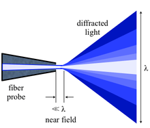

Near-field scanning optical microscopy (NSOM/SNOM) is a microscopy technique for nanostructure investigation that breaks the far field resolution limit by exploiting the properties of evanescent waves. This is done by placing the detector very close (distance much smaller than wavelength λ) to the specimen surface. This allows for the surface inspection with high spatial, spectral and temporal resolving power. With this technique, the resolution of the image is limited by the size of the detector aperture and not by the wavelength of the illuminating light. In particular, lateral resolution of 20 nm and vertical resolution of 2–5 nm have been demonstrated.[3][4] As in optical microscopy, the contrast mechanism can be easily adapted to study different properties, such as refractive index, chemical structure and local stress. Dynamic properties can also be studied at a sub-wavelength scale using this technique.
NSOM/SNOM is a form of scanning probe microscopy.
History
Edward Hutchinson Synge is given credit for conceiving and developing the idea for an imaging instrument that would image by exciting and collecting diffraction in the near field. His original idea, proposed in 1928, was based upon the usage of intense nearly planar light from an arc under pressure behind a thin, opaque metal film with a small orifice of about 100 nm. The orifice was to remain within 100 nm of the surface, and information was to be collected by point-by-point scanning. He foresaw the illumination and the detector movement being the biggest technical difficulties.[5][6] John A. O'Keefe also developed similar theories in 1956. He thought the moving of the pinhole or the detector when it is so close to the sample would be the most likely issue that could prevent the realization of such an instrument.[7][8] It was Ash and Nicholls who, in 1972, first broke the Abbe’s diffraction limit using radiation with wavelength of 3 cm. A line grating was resolved with a resolution of λ0/60.[9] A decade later, a patent on an optical near-field microscope was filed by Pohl,[10] followed in 1984 by the first paper that used visible radiation for near field scanning.[11] The near-field optical (NFO) microscope involved a subwavelength aperture at the apex of a metal coated sharply pointed transparent tip, and a feedback mechanism to maintain a constant distance of a few nanometers between the sample and the probe. Lewis et al. were also aware of the potential of an NFO microscope at this time.[12] They reported first results in 1986 confirming super-resolution.[13][14] In both experiments, details below 50 nm (about λ0/10) in size could be recognized.
Theory
According to Abbe’s theory of image formation, developed in 1873, the resolving capability of an optical component is ultimately limited by the spreading out of each image point due to diffraction. Unless the aperture of the optical component is large enough to collect all the diffracted light, the finer aspects of the image will not correspond exactly to the object. The minimum resolution (d) for the optical component are thus limited by its aperture size, and expressed by the Rayleigh criterion:

Here, λ0 is the wavelength in vacuum; NA is the numerical aperture for the optical component (maximum 1.3–1.4 for modern objectives with a very high magnification factor). Thus, the resolution limit is usually around λ0/2 for conventional optical microscopy.[15]
This treatment only assumes the light diffracted into the far-field that propagates without any restrictions. NSOM makes use of evanescent or non propagating fields that exist only near the surface of the object. These fields carry the high frequency spatial information about the object and have intensities that drop off exponentially with distance from the object. Because of this, the detector must be placed very close to the sample in the near field zone, typically a few nanometers. As a result, near field microscopy remains primarily a surface inspection technique. The detector is then rastered across the sample using a piezoelectric stage. The scanning can either be done at a constant height or with regulated height by using a feedback mechanism.[16]
Modes of operation
Aperture and apertureless operation

There exist NSOM which can be operated in so-called aperture mode and NSOM for operation in a non-aperture mode. As illustrated, the tips used in the apertureless mode are very sharp and do not have a metal coating.
Though there are many issues associated with the apertured tips (heating, artifacts, contrast, sensitivity, topology and interference amongst others), aperture mode remains more popular. This is primarily because apertureless mode is even more complex to set up and operate, and is not understood as well. There are five primary modes of apertured NSOM operation and four primary modes of apertureless NSOM operation. The major ones are illustrated in the next figure.


Some types of NSOM operation utilize a campanile probe, which has a square pyramid shape with two facets coated with a metal. Such a probe has a high signal collection efficiency (>90%) and no frequency cutoff.[19] Another alternative is “active tip" schemes, where the tip is functionalized with active light sources such as a fluorescent dye [20] or even a light emitting diode that enables fluorescence excitation.[21]
Feedback mechanisms
Feedback mechanisms are usually used to achieve high resolution and artifact free images since the detector must be positioned within a few nanometers of the surfaces. Some of these mechanisms are:
- Constant force feedback: This mode is very similar to the feedback mechanism used in atomic force microscopy (AFM). Experiments can be performed in contact, intermittent contact, and non-contact modes.
- Shear force feedback: In this mode, a tuning fork is mounted alongside the tip and made to oscillate at its resonance frequency. The amplitude is closely related to the tip-surface distance, and thus used as a feedback mechanism.[16]
Contrast
It is possible to take advantage of the various contrast techniques available to optical microscopy through NSOM but with much higher resolution. By using the change in the polarization of light or the intensity of the light as a function of the incident wavelength, it is possible to make use of contrast enhancing techniques such as staining, fluorescence, phase contrast and differential interference contrast. It is also possible to provide contrast using the change in refractive index, reflectivity, local stress and magnetic properties amongst others.[16][17]
Instrumentation and standard setup

The primary components of an NSOM setup are the light source, feedback mechanism, the scanning tip, the detector and the piezoelectric sample stage. The light source is usually a laser focused into an optical fiber through a polarizer, a beam splitter and a coupler. The polarizer and the beam splitter would serve to remove stray light from the returning reflected light. The scanning tip, depending upon the operation mode, is usually a pulled or stretched optical fiber coated with metal except at the tip or just a standard AFM cantilever with a hole in the center of the pyramidal tip. Standard optical detectors, such as avalanche photodiode, photomultiplier tube (PMT) or CCD, can be used. Highly specialized NSOM techniques, Raman NSOM for example, have much more stringent detector requirements.[17]
Near-field spectroscopy
As the name implies, information is collected by spectroscopic means instead of imaging in the near field regime. Through Near Field Spectroscopy (NFS), one can probe spectroscopically with subwavelength resolution. Raman SNOM and fluorescence SNOM are two of the most popular NFS techniques as they allow for the identification of nanosized features with chemical contrast. Some of the common near field spectroscopic techniques are:
- Direct local Raman NSOM: Aperture Raman NSOM is limited by very hot and blunt tips, and by long collection times. However, apertureless NSOM can be used to achieve high Raman scattering efficiency factors (around 40). Topological artifacts make it hard to implement this technique for rough surfaces.
- Tip-enhanced Raman spectroscopy (TERS) is an offshoot of Surface enhanced Raman spectroscopy (SERS). This technique can be used in an apertureless shear-force NSOM setup, or by using an AFM tip coated with gold or silver. The Raman signal is found to be significantly enhanced under the AFM tip. This technique has been used to give local variations in the Raman spectra under a single-walled nanotube. A highly sensitive optoacoustic spectrometer must be used for the detection of the Raman signal.
- Fluorescence NSOM: This highly popular and sensitive technique makes use of the fluorescence for near field imaging, and is especially suited for biological applications. The technique of choice here is the apertureless back to the fiber emission in constant shear force mode. This technique uses merocyanine based dyes embedded in an appropriate resin. Edge filters are used for removal of all primary laser light. Resolution as low as 10 nm can be achieved using this technique.
- Near field infrared spectrometry and near field dielectric microscopy:[17] near-field probes may be used to combine sub-micron microscopy with localised IR spectroscopy.[22]
Artifacts
NSOM is particularly vulnerable to artifacts that are not from the intended contrast mode. The most common root for artifacts in NSOM are:
- Tip breakage during scanning
- Striped contrast
- Displaced optical contrast
- Local far field light concentration
- Topographic artifacts
Limitations
- Very low working distance and extremely shallow depth of field.
- Limited to study surfaces.
- Not conducive for studying soft materials, especially under shear force mode.
- Long scan times for large sample areas for high resolution imaging.
See also
References
- ↑ Herzog, J. B. (2011). Optical Spectroscopy of Colloidal CdSe Semiconductor Nanostructures (PDF) (Ph.D. thesis). University of Notre Dame.
- ↑ Bao, Wei; Borys, Nicholas J.; Ko, Changhyun; Suh, Joonki; Fan, Wen; Thron, Andrew; Zhang, Yingjie; Buyanin, Alexander; Zhang, Jie; Cabrini, Stefano; Ashby, Paul D.; Weber-Bargioni, Alexander; Tongay, Sefaattin; Aloni, Shaul; Ogletree, D. Frank; Wu, Junqiao; Salmeron, Miquel B.; Schuck, P. James (2015). "Visualizing nanoscale excitonic relaxation properties of disordered edges and grain boundaries in monolayer molybdenum disulfide". Nature Communications. 6: 7993. Bibcode:2015NatCo...6E7993B. doi:10.1038/ncomms8993. PMC 4557266
 . PMID 26269394.
 - ↑ Dürig, U.; et al. (1986). "Near-field optical scanning microscopy". J. Appl. Phys. 59 (10): 3318. Bibcode:1986JAP....59.3318D. doi:10.1063/1.336848.
- ↑ Oshikane, Y.; et al. (2007). "Observation of nanostructure by scanning near-field optical microscope with small sphere probe". Sci. Technol. Adv. Mater. (free access). 8 (3): 181. Bibcode:2007STAdM...8..181O. doi:10.1016/j.stam.2007.02.013.
- ↑ Synge, E.H. (1928). "A suggested method for extending the microscopic resolution into the ultramicroscopic region". Phil. Mag. 6: 356. doi:10.1080/14786440808564615.
- ↑ Synge, E.H. (1932). "An application of piezoelectricity to microscopy". Phil. Mag. 13: 297. doi:10.1080/14786443209461931.
- ↑ O'Keefe, J.A. (1956). "Letters to the Editor". J. Opt. Soc. Am. 46: 359. Bibcode:1956JOSA...46..359.
- ↑ "Brief History and Simple Description of NSOM/SNOM Technology". Nanonics Inc. 12 October 2007.
- ↑ Ash, E.A. & Nicholls, G. (1972). "Super-resolution Aperture Scanning Microscope". Nature. 237 (5357): 510–2. Bibcode:1972Natur.237..510A. doi:10.1038/237510a0. PMID 12635200.
- ↑ EP patent 0112401, Pohl, Dieter Wolfgang, Dr., "optical near field scanning microscope", published 1987-04-22, issued 1982-12-27
- ↑ Pohl, D.W.; Denk, W. & Lanz, M. (1984). "Optical stethoscopy: Image recording with resolution λ/20". Appl. Phys. Lett. 44 (7): 651. Bibcode:1984ApPhL..44..651P. doi:10.1063/1.94865.
- ↑ Lewis, A.; Isaacson, M.; Harootunian, A. & Murray, A. (1984). "Development of a 500 Å spatial resolution light microscope. I. Light is efficiently transmitted through λ/16 diameter apertures". Ultramicroscopy. 13 (3): 227. doi:10.1016/0304-3991(84)90201-8.
- ↑ Betzig, E.; Lewis, A.; Harootunian, A.; Isaacson, M. & Kratschmer, E. (1986). "Near Field Scanning Optical Microscopy (NSOM)". Biophys. J. 49: 269. Bibcode:1986BpJ....49..269B. doi:10.1016/s0006-3495(86)83640-2.
- ↑ Harootunian, A.; Betzig, E.; Isaacson, M. & Lewis, A. (1986). "Super-resolution fluorescence near-field scanning optical microscopy". Appl. Phys. Lett. 49 (11): 674. Bibcode:1986ApPhL..49..674H. doi:10.1063/1.97565.
- ↑ Hecht, E. (2002). Optics. San Francisco: Addison Wesley. ISBN 0-19-510818-3.
- 1 2 3 Near-Field Scanning Optical Microscopy. Olympus America Inc. 12 October 2007.
- 1 2 3 4 5 Kaupp, G. (2006). Atomic Force Microscopy, Scanning Nearfield Optical Microscopy and Nanoscratching: Application to Rough and Natural Surfaces. Heidelberg: Springer. ISBN 3-540-28405-2.
- 1 2 Introduction to NSOM. The Optics Laboratory, North Carolina State University. 12 October 2007
- ↑ Bao, W.; Melli, M.; Caselli, N.; Riboli, F.; Wiersma, D. S.; Staffaroni, M.; Choo, H.; Ogletree, D. F.; Aloni, S.; Bokor, J.; Cabrini, S.; Intonti, F.; Salmeron, M. B.; Yablonovitch, E.; Schuck, P. J.; Weber-Bargioni, A. (2012). "Mapping Local Charge Recombination Heterogeneity by Multidimensional Nanospectroscopic Imaging". Science. 338 (6112): 1317. Bibcode:2012Sci...338.1317B. doi:10.1126/science.1227977. PMID 23224550.
- ↑ Sandoghdar, V.; Michaelis, J.; Hettich, C.; Mlynek, J. (2000). "Optical microscopy using a single-molecule light source". Nature. 405 (6784): 325. doi:10.1038/35012545.
- ↑ Hoshino, Kazunori; Gopal, Ashwini; Glaz, Micah S.; Vanden Bout, David A.; Zhang, Xiaojing (2012). "Nanoscale fluorescence imaging with quantum dot near-field electroluminescence". Applied Physics Letters. 101 (4): 043118. Bibcode:2012ApPhL.101d3118H. doi:10.1063/1.4739235.
- ↑ H M Pollock & D A Smith (2002). "The use of near-field probes for vibrational spectroscopy and photothermal imaging". In J M Chalmers & P R Griffiths. Handbook of vibrational spectroscopy vol. 2. pp. 1472–92.
External links
- SNOM Scan Image Gallery at the Wayback Machine (archived October 2, 2008)

Optical microscopy | ||
|---|---|---|
| Illumination and contrast methods |  | |
| Fluorescence methods | ||
| Sub-diffraction limit techniques | ||