Dorsal scapular nerve
| Dorsal scapular nerve | |
|---|---|
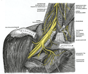 The right brachial plexus with its short branches, viewed from in front. (Dorsalis scapulae labeled at left, second from top.) | |
| Details | |
| From | C5 of brachial plexus |
| Innervates | rhomboid minor, rhomboid major, levator scapulae |
| Identifiers | |
| Latin | nervus dorsalis scapulae |
| TA | A14.2.03.011 |
| FMA | 65279 |
The dorsal scapular nerve arises from the brachial plexus, usually from the plexus root (anterior/ventral ramus) of the cervical nerve C5. Once the nerve leaves C5 it commonly pierces the middle scalene muscle, and continues deep to levator scapulae and the rhomboids (minor superior to major).
It provides motor innervation to the rhomboid muscles, which pull the scapula towards the spine and levator scapulae muscle, which elevates the scapula.
Injury to this nerve is usually apparent on inspection when the scapula on the injured side is located farther from the midline than the uninjured scapula. The patient would be unable to pull their shoulder back, as when standing at attention. Isolated dorsal scapular nerve injury is uncommon, but case reports usually involve injury to the scalene muscles. [1]
The nerve is accompanied by one of two arteries: either the dorsal scapular artery (the only artery that branches off the third part of the subclavian artery, although its origin is highly variable in humans) or, when the dorsal scapular artery is absent, the deep branch of the transverse cervical artery. The latter is one of three arteries branching off the thyrocervical trunk, a branch of the first part of the subclavian artery, with the other two branches being the vertebral artery and internal thoracic artery.
See also
- Dorsal scapular artery
- Not to be confused with: thoracodorsal nerve
Additional images
-

Brachial plexus
-
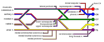
Brachial plexus with courses of spinal nerves shown
-

Deep Branch of Transverse Cervical running with Dorsal Scapular
External links
- Dorsal_scapular_nerve at the Duke University Health System's Orthopedics program