Rhomboid minor muscle
| Rhomboid minor | |
|---|---|
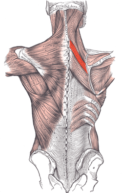 Muscles connecting the upper extremity to the vertebral column. (Rhomboid minor in red) | |
| Details | |
| Origin | Nuchal ligaments and spinous processes of C7-T1 |
| Insertion | Medial border of scapula, superior to the insertion of rhomboid major muscle |
| Artery | Deep branch of transverse cervical artery |
| Nerve | Dorsal scapular nerve (C4–5) |
| Actions | Retracts and rotates scapula, fixes scapula to thoracic wall |
| Antagonist | Serratus anterior |
| Identifiers | |
| Latin | Musculus rhomboideus minor |
| TA | A04.3.01.008 |
| FMA | 13380 |
In human anatomy, the rhomboid minor is a small skeletal muscle on the back that connects the scapula with the vertebrae of the spinal column.
Located inferior to levator scapulae and superior to rhomboid major, it acts together with the latter to keep the scapula pressed against the thoracic wall. It lies deep to trapezius but superficial to the long spinal muscles.[1]
Origin and insertion
The rhomboid minor arises from the inferior border of the nuchal ligament, from the spinous processes of the seventh cervical and first thoracic vertebrae, and from the intervening supraspinous ligaments.[1]
It is inserted into a small area of the medial border of the scapula at the level of the scapular spine.[2]
Action
Together with the rhomboid major, the rhomboid minor retracts the scapula when trapezius is contracted. Acting as an antagonist to the trapezius, the rhomboid major and minor elevate the medial border of the scapula medially and upward, working in tandem with the levator scapulae muscle to rotate the scapulae downward. While other shoulder muscles are active, the rhomboid major and minor stabilize the scapula. [3]
Innervation and blood supply
The nerve supply comes from the dorsal scapular nerve, with most of its fibers derived from the C5 nerve root and only minor contribution from C4 or C6. [4]
The rhomboid minor gets its arterial blood supply from the dorsal scapular artery.
Variation
It is usually separated from the rhomboid major by a slight interval, but the adjacent margins of the two muscles are occasionally united. [5]
Additional images
|


References
This article incorporates text in the public domain from the 20th edition of Gray's Anatomy (1918)
- 1 2 194641998 at GPnotebook
- ↑ Origin, insertion and nerve supply of the muscle at Loyola University Chicago Stritch School of Medicine
- ↑ "Function (of rhomboid muscles)". GP Notebook. Retrieved January 2011. Check date values in:
|access-date=(help) - ↑ Martin, R. M.; Fish, D. E. (2007). "Scapular winging: anatomical review, diagnosis, and treatments". Current Reviews in Musculoskeletal Medicine. 1 (1): 1–11. doi:10.1007/s12178-007-9000-5. PMC 2684151
 . PMID 19468892., p. 4
 - ↑ Gray's Anatomy (1918), see infobox
External links
| Wikimedia Commons has media related to Rhomboid minor muscles. |
- Anatomy photo:01:st-0211 at the SUNY Downstate Medical Center