Vertebral artery
| Vertebral artery | |
|---|---|
 The vertebral arteries arise from the subclavian arteries, unite as the basilar artery, and supplies blood to parts of the brain | |
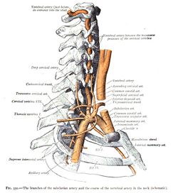 The branches of the subclavian artery and the course of the vertebral artery in the neck (schematic). | |
| Details | |
| Source | subclavian arteries |
| Branches |
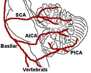
Meningeal branches Posterior spinal Anterior spinal PICA Basilar artery |
| Vein | vertebral vein |
| Supplies | Upper spinal cord, brainstem, cerebellum, posterior part of brain |
| Identifiers | |
| Latin | arteria vertebralis |
| MeSH | A07.231.114.839 |
| TA | A12.2.08.002 |
| FMA | 3956 |
The vertebral arteries are major arteries of the neck. They arise as branches from the subclavian arteries and merge to form the single midline basilar artery. As the vertebrobasilar system, they supply blood to the upper spinal cord, brainstem, cerebellum, and posterior part of brain.
Structure

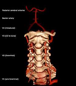
The vertebral arteries arise from the subclavian arteries, one on each side of the body, then enter deep to the transverse process at the level of the 6th cervical vertebrae (C6),[1] or occasionally (in 7.5% of cases) at the level of C7. They then proceed superiorly, in the transverse foramen of each cervical vertebra.[1] Once they have passed through the transverse foramen of C1 (also known as the atlas), the vertebral arteries travel across the posterior arch of C1 and through the suboccipital triangle before entering the foramen magnum.[1]
Nunziante Ippolito, a Neapolitan physician, identified the "angle of Nunziante Ippolito" to find the vertebral artery, between the anterior scalene muscle and the longus colli muscle.[2]
Inside the skull, the two vertebral arteries join to form the basilar artery at the base of the Pons. The basilar artery is the main blood supply to the brainstem and connects to the Circle of Willis to potentially supply the rest of the brain if there is compromise to one of the carotids. At each cervical level, the vertebral artery sends branches to the surrounding musculature via the anterior spinal arteries.
The vertebral artery may be divided into four parts:
- The first part runs upward and backward between the Longus colli and the Scalenus anterior. In front of it are the internal jugular and vertebral veins, and it is crossed by the inferior thyroid artery; the left vertebral is crossed by the thoracic duct also. Behind it are the transverse process of the seventh cervical vertebra, the sympathetic trunk and its inferior cervical ganglion
- The second part runs upward through the foramina in the transverse processes of the C6 to C2 vertebræ, and is surrounded by branches from the inferior cervical sympathetic ganglion and by a plexus of veins which unite to form the vertebral vein at the lower part of the neck. It is situated in front of the trunks of the cervical nerves, and pursues an almost vertical course as far as the transverse process of the axis.
- The third part issues from the C2 foramen transversarium on the medial side of the Rectus capitis lateralis. It is further subdivided into the vertical part V3v passing vertically upwards, crossing the C2 root and entering the foramen transversarium of C1, and the horizontal part V3h, curving medially and posteriorly behind the superior articular process of the atlas, the anterior ramus of the first cervical nerve being on its medial side; it then lies in the groove on the upper surface of the posterior arch of the atlas, and enters the vertebral canal by passing beneath the posterior atlantoöccipital membrane. This part of the artery is covered by the Semispinalis capitis and is contained in the suboccipital triangle—a triangular space bounded by the Rectus capitis posterior major, the Obliquus superior, and the Obliquus inferior. The first cervical or suboccipital nerve lies between the artery and the posterior arch of the atlas.
- The fourth part pierces the dura mater and inclines medialward to the front of the medulla oblongata; it is placed between the hypoglossal nerve and the anterior root of the first cervical nerve and beneath the first digitation of the ligamentum denticulatum. At the lower border of the pons it unites with the vessel of the opposite side to form the basilar artery.
Variation
The left vertebral artery is usually larger and carries more blood.[3] In 3-15% of the population, a bony bridge called the arcuate foramen covers the groove for the vertebral artery on vertebra C1.
Function
Upper spinal cord, brainstem, cerebellum, posterior part of brain.[1]
References
- 1 2 3 4 editor-in-chief, Susan Standring ; section editors, Neil R. Borley; et al. (2008). Gray's anatomy : the anatomical basis of clinical practice (40th ed.). London: Churchill Livingstone. ISBN 978-0-8089-2371-8.
- ↑ http://www.treccani.it/enciclopedia/nunziante-ippolito_%28Dizionario_Biografico%29/[]
- ↑ Albayrak, Ramazan; Degirmenci, B; Acar, M; Haktanir, A; Colbay, M; Yaman, M (2007). "Doppler sonography evaluation of flow velocity and volume of the extracranial internal carotid and vertebral arteries in healthy adults". J Clin Ultrasound. 35 (1): 27–33. doi:10.1002/jcu.20301. PMID 17149761.
Additional images
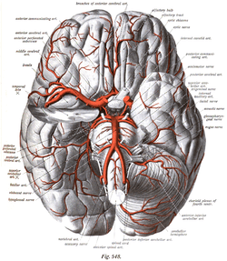 The arteries of the base of the brain.
 Diagram of the arterial circulation at the base of the brain.
 Relationship of the vertebral artery to the suboccipital muscles.
External links
| Wikimedia Commons has media related to Vertebral artery. |
- Anatomy photo:28:09-0201 at the SUNY Downstate Medical Center
- http://neuroangio.org/anatomy-and-variants/vertebral-artery/
- MedEd at Loyola Neuro/neurovasc/navigation/vert.htm
- Atlas image: n3a8p1 at the University of Michigan Health System
- lesson5 at The Anatomy Lesson by Wesley Norman (Georgetown University) (vertebralcolumnfromleftweb)
- Anatomy diagram: 13048.000-1 at Roche Lexicon - illustrated navigator, Elsevier
- Anatomy diagram: 13048.000-3 at Roche Lexicon - illustrated navigator, Elsevier