Armillaria mellea
| Armillaria mellea | |
|---|---|
 | |
| Scientific classification | |
| Kingdom: | Fungi |
| Division: | Basidiomycota |
| Class: | Agaricomycetes |
| Order: | Agaricales |
| Family: | Physalacriaceae |
| Genus: | Armillaria |
| Species: | A. mellea |
| Binomial name | |
| Armillaria mellea (Vahl) P.Kumm. (1871) | |
| Synonyms[1] | |
| |
| Armillaria mellea | |
|---|---|
|
| |
| gills on hymenium | |
|
cap is convex or flat | |
|
hymenium is adnate or subdecurrent | |
| stipe has a ring | |
| spore print is white | |
| ecology is parasitic | |
|
edibility: edible but not recommended | |
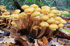
Armillaria mellea, commonly known as honey fungus, is a basidiomycete fungus in the genus Armillaria. It is a plant pathogen and part of a cryptic species complex of closely related and morphologically similar species. It causes Armillaria root rot in many plant species and produces mushrooms around the base of trees it has infected. The symptoms of infection appear in the crowns of infected trees as discoloured foliage, reduced growth, dieback of the branches and death. The mushrooms are edible but some people may be intolerant to them. This species is capable of producing light via bioluminescence in its mycelium.
Armillaria mellea is widely distributed in temperate regions of the Northern Hemisphere. The fruit body or mushroom, commonly known as stump mushroom, stumpie, honey mushroom, pipinky or pinky, grows typically on hardwoods but may be found around and on other living and dead wood or in open areas.
Taxonomy
The species was originally named Agaricus melleus by Danish-Norwegian botanist Martin Vahl in 1790; it was transferred to the genus Armillaria in 1871 by Paul Kummer.[1] Numerous subtaxa have been described:
| Name | Authority | Year |
|---|---|---|
| var. viridiflava | Barla[2] | 1887 |
| var. minor | Barla[2] | 1887 |
| var. bulbosa | Barla[2] | 1887 |
| var. camerunensis | Henn.[3] | 1895 |
| var. exannulata | Peck[4] | 1893 |
| var. flava | Peck[5] | 1897 |
| var. glabra | Gillet[6] | 1874 |
| var. javanica | Henn.[7] | 1900 |
| var. laricina | (Bolton) Barla[2] | 1887 |
| var. maxima | Barla[2] | 1887 |
| var. obscura | Gillet[6] | 1874 |
| var. radicata | Peck[8] | 1891 |
| var. sulphurea | (Weinm.) Fr.[9] | 1879 |
| var. tabescens | (Scop.) Rea & Ramsb. | 1917 |
| var. versicolor | (With.) W.G.Sm.[10] | 1908 |
| subsp. nipponica | J.Y.Cha & Igarashi[11] | 1995 |
| f. rosea | Calonge & M.Seq.[12] | 2003 |
Description

The basidiocarp of each has a smooth cap 3 to 15 cm (1 to 6 in) in diameter, convex at first but becoming flattened with age often with a central raised umbo, later becoming somewhat dish-shaped. The margins of the cap are often arched at maturity and the surface is sticky when wet. Though typically honey-coloured, this fungus is rather variable in appearance and sometimes has a few dark, hairy scales near the centre somewhat radially arranged. The gills are white at first, sometimes becoming pinkish-yellow or discoloured with age, broad and fairly distant, attached to the stipe at right angles or are slightly decurrent. The spore print is white. The stipe is of variable length, up to about 20 cm (8 in) long and 3.5 cm (1.4 in) in diameter. It is fibrillose and of a firm spongy consistency at first but later becomes hollow. It is cylindrical and tapers to a point at its base where it is fused to the stipes of other mushrooms in the clump. It is whitish at the upper end and brownish-yellow below, often with a very dark-coloured base. There is a broad persistent skin-like ring attached to the upper part of the stipe. This has a velvety margin and yellowish fluff underneath and extends outwards as a white partial veil protecting the gills when young. The flesh of the cap is whitish and has a sweetish odour and flavour with a tinge of bitterness. Under the microscope, the spores are approximately elliptical, 7–9 by 6–7 µm, inamyloid with prominent apiculi (short, pointed projections) at the base. The basidia (spore-producing structures) lack basal clamps.[13][14]
The main part of the fungus is underground where a mat of mycelial threads may extend for great distances. They are bundled together in rhizomorphs that are black in this species.[14] The fungal body is not bioluminescent but its mycelia are luminous when in active growth.[15]
Similar species
Armillaria mellea once included a range of species with similar features that have since been reclassified.[16]
Distribution
Armillaria mellea is widespread in northern temperate zones. It has been found in North America, Europe and northern Asia, and It has been introduced to South Africa. The fungus grows parasitically on a large number of broadleaf trees. It fruits in dense clusters at the base of trunks or stumps.[17]
Ecology
Trees become infected by Armillaria mellea when rhizomorphs growing through the soil encounter uninfected roots. Alternatively, when infected roots come into contact with uninfected ones the fungal mycelium may grow across. The rhizomorphs invade the trunk, growing between the bark and the wood and causing wood decay, growth reduction and mortality. Trees that are already under stress are more likely to be attacked but healthy trees may also be parasitized. The foliage becomes sparse and discoloured, twig growth slows down and branches may die back. When they are attacked, the Douglas-fir, western larch and some other conifers often produce an extra large crop of cones shortly before dying. Coniferous trees also tend to ooze resin from infected areas whereas broad-leaved trees sometimes develop sunken cankers. A growth of fruiting bodies near the base of the trunk confirms the suspicion of Armillaria root rot.[18]
In 1893, the American mycologist Charles Horton Peck reported finding Armillaria fruiting bodies that were "aborted", in a similar way to specimens of Entoloma abortivum. It was not until 1974 that Roy Watling showed that the aborted specimens included cells of both Armillaria mellea and Entoloma abortivum. He thought that the Armillaria was parasitizing the Entoloma, a plausible hypothesis given its pathogenic behaviour.[19] However, a 2001 study by Czederpiltz, Volk and Burdsall showed that the Entoloma was in fact the microparasite. The whitish-grey malformed fruit bodies known as carpophoroids were the result of E. abortivum hyphae penetrating the Armillaria and disrupting its normal development.[20]
The main part of the fungus is underground where a mat of mycelial threads may extend for great distances. The rhizomorphs of A. mellea are initiated from mycelium into multicellular apices of rhizomorphs, which are multicellular vegetative organs that exclude soil from the interior of the rhizomorph tissues. The rhizomorphs spread through far greater distances through the ground than the mycelium. The rhizomorphs are black in this species.[14] The fungal body is not bioluminescent but its mycelia and rhizomorphs are luminous when in active growth.[15] A. mellea producing rhizomorphs is parasitic on woody plants of many species, including especially shrubs, hardwood and evergreen trees. In one example, spread by rhizomorphs from an initially infected tree killed 600 trees in a prune orchard in 6 years. Each infected tree was immediately adjacent to an already infected one, the spread by rhizomorphs through the tree roots and soil. (Piper and Fletcher, 1903, Wash. Age. Exp. Sat. But., 59: 1-14); cited in Rhizomorph Development in A. mellea, Ph.D. thesis, by Philip Snider(1957), Farlow Herbarium Library Harvard Univ., 20 Divinity Ave., Cambridge, Mass.
Edibility
Armillaria mellea mushroom are considered good edibles, although some individuals have reported "allergic" reactions that result in stomach upsets. Some authors suggest not collecting mushrooms from the wood of various trees, including hemlock, buckeye, eucalyptus, and locust. The mushrooms have a taste that has been described as slightly sweet and nutty, with a texture ranging from chewy to crunchy, depending on the method of preparation. Parboiling mushrooms before consuming removes the bitter taste present in some specimens, and may reduce the amount of gastrointestinal irritants. Drying the mushrooms preserves and intensifies their flavour, although reconstituted mushrooms tend to be tough to eat.[21]
Chemistry
Several bioactive compounds have been isolated and identified from the fruit bodies. The triterpenes 3β-hydroxyglutin-5-ene, friedelane-2α,3β-diol, and friedelin were reported in 2011.[22] Indole compounds include tryptamine, L-tryptophan and serotonin.[23]
See also
References
- 1 2 "Armillaria mellea (Vahl) P. Kumm., Der Führer in die Pilzkunde: 134, 1871". MycoBank. International Mycological Association. Retrieved 2013-10-19.
- 1 2 3 4 5 Barla JB. (1887). Liste des champignons nouvellement observés dans le département des Alpes-Maritimes. Bulletin de la Société Mycologique de France (in French). 3. pp. 138–44.
- ↑ Hennings P. (1895). "Fungi camerunenses I". Botanische Jahrbücher für Systematik Pflanzengeschichte und Pflanzengeographie (in German). 22: 72–111 (see p. 107).
- ↑ Peck CH. (1893). "Report of the Botanist (1892)". Annual Report on the New York State Museum of Natural History. 46: 85–149 (see p. 134).
- ↑ Peck CH. (1896). "Report of the Botanist (1894)". Annual Report on the New York State Museum of Natural History. 48: 103–337 (see p. 265).
- 1 2 Gillet CC. (1874). Les Hyménomycètes ou Description de tous les Champignons qui Croissent en France (in French). 1. Alençon: Ch. Thomas. p. 84.
- ↑ Hennings P. (1900). "Fungi monsunenses". Monsunia. 1: 1–38.
- ↑ Peck CH. (1891). "Report of the Botanist (1890)". Annual Report on the New York State Museum of Natural History. 44: 117–87 (see p. 150).
- ↑ Karsten PA. (1879). "Rysslands, Finlands och den Skandinaviska halföns Hattsvampar. Förra Delen: Skifsvampar". Bidrag till Kännedom av Finlands Natur och Folk (in German). 32: 22.
- ↑ Smith WG. (1908). Synopsis of the British Basidiomycetes: A descriptive catalogue of the drawings and specimens in the department of Botany British Museum. London, UK: The Trustees of the British Museum, London. p. 30.
- ↑ Cha JY, Igarashi T (1995). "A note on Armillaria mellea subsp. nipponica subsp. nov. in Japan". Mycoscience. 36 (2): 143–6. doi:10.1007/BF02268548.
- ↑ Calonge FD, Menezes de Sequeira M (2003). "Contribución al catálogo de los hongos de Madeira (Portugal)". Boletín de la Sociedad Micológica de Madrid (in Spanish). 27: 277–308.
- ↑ Hvass, Else; Hvass, Hans (1961). Mushrooms and Toadstools in Colour. Blandford Press. p. 110. ISBN 9780713701463.
- 1 2 3 Kuo, Michael (2004-10-01). "Armillaria mellea: The Honey Mushroom". MushroomExpert.Com. Retrieved 2013-10-18.
- 1 2 Desjardin DE, Oliveira AG, Stevani CV (2008). "Fungi bioluminescence revisited". Photochemical & Photobiological sciences. 7 (2): 170–82. doi:10.1039/b713328f. PMID 18264584.
- ↑ Ross-Davis AL, Hanna JW, Kim MS, Klopfenstein NB (2012). "Advances toward DNA-based identification and phylogeny of North American Armillariaspecies using elongation factor-1 alpha gene". Mycoscience. 53 (2): 161–5. doi:10.1007/s10267-011-0148-x.
- ↑ Roberts P, Evans S (2011). The Book of Fungi. Chicago, Illinois: University of Chicago Press. p. 63. ISBN 978-0-226-72117-0.
- ↑ Williams, RE; Shaw, CG; Wargo, PM; Sites, WH (1989-04-01). "Armillaria Root Disease". Forest Insect & Disease Leaflet 78. US Department of Agriculture Forest Service. Retrieved 2013-10-17.
- ↑ Kuo, Michael (2004-10-01). "Entoloma abortivum". MushroomExpert.Com. Retrieved 2013-10-19.
- ↑ Czederpiltz DL, Volk TJ, Burdsall HH Jr (2001). "Field observations and inoculation experiments to determine the nature of the carpophoroids associated with Entoloma abortivum and Armillaria". Mycologia. 93 (5): 841–51. doi:10.2307/3761750. JSTOR 3761750.
- ↑ Kuo M. (2007). 100 Edible Mushrooms. Ann Arbor, Michigan: The University of Michigan Press. pp. 244–6. ISBN 978-0-472-03126-9.
- ↑ Guo WJ, Guo SX (2011). "Triterpene from Armillaria mellea". Chemistry of Natural Compounds. 46 (6): 995–6. doi:10.1007/s10600-011-9809-4.
- ↑ Muszynska B, Maslanka A, Ekiert H, Sulkowska-Ziaja K (2011). "Analysis of indole compounds in Armillaria mellea fruiting bodies". Acta Poloniae Pharmaceutica. 68 (1): 93–7. PMID 21485706.
External links
| Wikimedia Commons has media related to Armillaria mellea. |