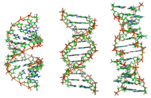
Z-DNA

Z-DNA is one of the many possible double helical structures of DNA. It is a left-handed double helical structure in which the double helix winds to the left in a zig-zag pattern (instead of to the right, like the more common B-DNA form). Z-DNA is thought to be one of three biologically active double helical structures along with A- and B-DNA.
History
Left handed DNA was first discovered by Robert Wells and colleagues, during their studies of a repeating polymer of inosine-cytosine.[1] They observed a "reverse" circular dichroism spectrum for such DNAs, and interpreted this (correctly) to mean that the strands wrapped around one another in a left handed fashion. The relationship between Z-DNA and the more familiar B-DNA was indicated by the work of Pohl and Jovin,[2] who showed that the ultraviolet circular dichroism of poly(dG-dC) was nearly inverted in 4 M sodium chloride solution. The suspicion that this was the result of a conversion from B-DNA to Z-DNA was confirmed by examining the raman spectra of these solutions and the Z-DNA crystals.[3] Subsequently, a crystal structure of "Z-DNA" was published which turned out to be the first single-crystal X-ray structure of a DNA fragment (a self-complementary DNA hexamer d(CG)3). It was resolved as a left-handed double helix with two anti-parallel chains that were held together by Watson-Crick base pairs (see: x-ray crystallography). It was solved by Andrew Wang, Alexander Rich, and co-workers in 1979 at MIT.[4] The crystallisation of a B- to Z-DNA junction in 2005[5] provided a better understanding of the potential role Z-DNA plays in cells. Whenever a segment of Z-DNA forms, there must be B-Z junctions at its two ends, interfacing it to the B-form of DNA found in the rest of the genome.
In 2007, the RNA version of Z-DNA, Z-RNA, was described as a transformed version of an A-RNA double helix into a left-handed helix.[6] The transition from A-RNA to Z-RNA, however, was already described in 1984.[7]
Structure

Z-DNA is quite different from the right-handed forms. In fact, Z-DNA is often compared against B-DNA in order to illustrate the major differences. The Z-DNA helix is left-handed and has a structure that repeats every 2 base pairs. The major and minor grooves, unlike A- and B-DNA, show little difference in width. Formation of this structure is generally unfavourable, although certain conditions can promote it; such as alternating purine-pyrimidine sequence (especially poly(dGC)2), negative DNA supercoiling or high salt and some cations (all at physiological temperature, 37 °C, and pH 7.3-7.4). Z-DNA can form a junction with B-DNA (called a "B-to-Z junction box") in a structure which involves the extrusion of a base pair.[8] The Z-DNA conformation has been difficult to study because it does not exist as a stable feature of the double helix. Instead, it is a transient structure that is occasionally induced by biological activity and then quickly disappears.[9]
Predicting Z-DNA structure
It is possible to predict the likelihood of a DNA sequence forming a Z-DNA structure. An algorithm for predicting the propensity of DNA to flip from the B-form to the Z-form, ZHunt, was written by Dr. P. Shing Ho in 1984 (at MIT).[10] This algorithm was later developed by Tracy Camp, P. Christoph Champ, Sandor Maurice, and Jeffrey M. Vargason for genome-wide mapping of Z-DNA (with P. Shing Ho as the principal investigator).[11]
Biological significance
While no definitive biological significance of Z-DNA has been found, it is commonly believed to provide torsional strain relief (supercoiling) while DNA transcription occurs.[5][12] The potential to form a Z-DNA structure also correlates with regions of active transcription. A comparison of regions with a high sequence-dependent, predicted propensity to form Z-DNA in human chromosome 22 with a selected set of known gene transcription sites suggests there is a correlation.[11]
Toxic effect of ethidium bromide on trypanosomas is caused by shift of their kinetoplastid DNA to Z-form. The shift is caused by intercalation of EtBr and subsequent loosening of DNA structure that leads to unwinding of DNA, shift to Z-form and inhibition of DNA replication.[13]
Z-DNA formed after transcription initiation
The first domain to bind Z-DNA with high affinity was discovered in ADAR1 using an approach developed by Alan Herbert.[14][15] Crystallographic and NMR studies confirmed the biochemical findings that this domain bound Z-DNA in a non-sequence-specific manner.[16][17][18] Related domains were identified in a number of other proteins through sequence homology.[15] The identification of the Z-alpha domain provided a tool for other crystallographic studies that lead to the characterization of Z-RNA and the B-Z junction. Biological studies suggested that the Z-DNA binding domain of ADAR1 may localize this enzyme that modifies the sequence of the newly formed RNA to sites of active transcription.[19][20]
In 2003, Alex Rich noticed that a poxvirus virulence factor, called E3L that has a Z-alpha related domain, mimicked a mammalian protein that binds Z-DNA.[21][22] In 2005, Rich and his colleagues pinned down what E3L does for the poxvirus. When expressed in human cells, E3L increases by five- to 10-fold the production of several genes that block a cell's ability to self-destruct in response to infection.
Rich speculates that the Z-DNA is necessary for transcription and that E3L stabilizes the Z-DNA, thus prolonging expression of the anti-apoptotic genes. He suggests that a small molecule that interferes with the E3L binding to Z-DNA could thwart the activation of these genes and help protect people from pox infections.
Comparison geometries of some DNA forms

| Geometry attribute | A-form | B-form | Z-form |
|---|---|---|---|
| Helix sense | right-handed | right-handed | left-handed |
| Repeating unit | 1 bp | 1 bp | 2 bp |
| Rotation/bp | 32.7° | 34.3° | 60°/2 |
| bp/turn | 11 | 10 | 12 |
| Inclination of bp to axis | +19° | −1.2° | −9° |
| Rise/bp along axis | 2.3 Å (0.23 nm) | 3.32 Å (0.332 nm) | 3.8 Å (0.38 nm) |
| Pitch/turn of helix | 28.2 Å (2.82 nm) | 33.2 Å (3.32 nm) | 45.6 Å (4.56 nm) |
| Mean propeller twist | +18° | +16° | 0° |
| Glycosyl angle | anti | anti | C: anti, G: syn |
| Sugar pucker | C3'-endo | C2'-endo | C: C2'-endo, G: C3'-endo |
| Diameter | 23 Å (2.3 nm) | 20 Å (2.0 nm) | 18 Å (1.8 nm) |
| Sources:[23][24][25] | |||
See also
References
- ↑ Mitsui; et al. (1970). "Physical and enzymatic studies on poly d(I-C)-poly d(I-C), an unusual double-helical DNA". Nature (London). 228 (5277): 1166–1169. doi:10.1038/2281166a0. PMID 4321098.
- ↑ Pohl FM, Jovin TM (1972). "Salt-induced co-operative conformational change of a synthetic DNA: equilibrium and kinetic studies with poly(dG-dC)". J. Mol. Biol. 67 (3): 375–396. doi:10.1016/0022-2836(72)90457-3. PMID 5045303.
- ↑ Thamann TJ, Lord RC, Wang AH, Rich A (1981). "High salt form of poly(dG-dC)•poly(dG-dC) is left handed Z-DNA: raman spectra of crystals and solutions". Nucl. Acids Res. 9 (20): 5443–5457. doi:10.1093/nar/9.20.5443. PMC 327531
 . PMID 7301594.
 - ↑ Wang AH, Quigley GJ, Kolpak FJ, Crawford JL, van Boom JH, Van der Marel G, Rich A (1979). "Molecular structure of a left-handed double helical DNA fragment at atomic resolution". Nature (London). 282 (5740): 680–686. Bibcode:1979Natur.282..680W. doi:10.1038/282680a0. PMID 514347.
- 1 2 Ha SC, Lowenhaupt K, Rich A, Kim YG, Kim KK (2005). "Crystal structure of a junction between B-DNA and Z-DNA reveals two extruded bases". Nature. 437 (7062): 1183–1186. Bibcode:2005Natur.437.1183H. doi:10.1038/nature04088. PMID 16237447.
- ↑ Placido D, Brown BA 2nd, Lowenhaupt K, Rich A, Athanasiadis A (2007). "A left-handed RNA double helix bound by the Zalpha domain of the RNA-editing enzyme ADAR1". Structure. 15 (4): 395–404. doi:10.1016/j.str.2007.03.001. PMC 2082211. PMID 17437712.
- ↑ Hall K, Cruz P, Tinoco I Jr, Jovin TM, van de Sande JH (October 1984). "'Z-RNA'--a left-handed RNA double helix". Nature. 311 (5986): 584–586. Bibcode:1984Natur.311..584H. doi:10.1038/311584a0. PMID 6482970.
- ↑ de Rosa M, de Sanctis D, Rosario AL, Archer M, Rich A, Athanasiadis A, Carrondo MA (2010-05-18). "Crystal structure of a junction between two Z-DNA helices". Proc Natl Acad Sci USA. 107 (20): 9088–9092. Bibcode:2010PNAS..107.9088D. doi:10.1073/pnas.1003182107. PMC 2889044. PMID 20439751.
- ↑ Zhang H, Yu H, Ren J, Qu X (2006). "Reversible B/Z-DNA transition under the low salt condition and non-B-form polydApolydT selectivity by a cubane-like europium-L-aspartic acid complex". Biophysical Journal. 90 (9): 3203–3207. Bibcode:2006BpJ....90.3203Z. doi:10.1529/biophysj.105.078402. PMC 1432110. PMID 16473901.
- ↑ Ho PS, Ellison MJ, Quigley GJ, Rich A (1986). "A computer aided thermodynamic approach for predicting the formation of Z-DNA in naturally occurring sequences". EMBO Journal. 5 (10): 2737–2744. PMC 1167176. PMID 3780676.
- 1 2 Champ PC, Maurice S, Vargason JM, Camp T, Ho PS (2004). "Distributions of Z-DNA and nuclear factor I in human chromosome 22: a model for coupled transcriptional regulation". Nucleic Acids Res. 32 (22): 6501–6510. doi:10.1093/nar/gkh988. PMC 545456. PMID 15598822.
- ↑ Rich A, Zhang S (2003). "Timeline: Z-DNA: the long road to biological function". Nature Reviews Genetics. 4 (7): 566–572. doi:10.1038/nrg1115. PMID 12838348.
- ↑ Roy Chowdhury, Arnab; Bakshi, Rahul; Wang, Jianyang; Yildirir, Gokben; Liu, Beiyu; Pappas-Brown, Valeria; Tolun, Gökhan; Griffith, Jack D.; Shapiro, Theresa A.; Jensen, Robert E.; Englund, Paul T.; Ullu, Elisabetta (16 December 2010). "The Killing of African Trypanosomes by Ethidium Bromide". PLoS Pathogens. 6 (12): e1001226. doi:10.1371/journal.ppat.1001226.
- ↑ Herbert A, Rich A (1993). "A method to identify and characterize Z-DNA binding proteins using a linear oligodeoxynucleotide". Nucleic Acids Res. 21 (11): 2669–72. doi:10.1093/nar/21.11.2669. PMC 309597. PMID 8332463.
- 1 2 Herbert A, Alfken J, Kim YG, Mian IS, Nishikura K, Rich A (1997). "A Z-DNA binding domain present in the human editing enzyme, double-stranded RNA adenosine deaminase.". Proc Natl Acad Sci USA. 94 (16): 8421–6. Bibcode:1997PNAS...94.8421H. doi:10.1073/pnas.94.16.8421. PMC 22942. PMID 9237992.
- ↑ Herbert A, Schade M, Lowenhaupt K, Alfken J, Schwartz T, Shlyakhtenko LS, Lyubchenko YL, Rich A (1998). "The Zalpha domain from human ADAR1 binds to the Z-DNA conformer of many different sequences". Nucleic Acids Res. 26 (15): 2669–72. doi:10.1093/nar/26.15.3486. PMC 147729. PMID 9671809.
- ↑ Schwartz T, Rould MA, Lowenhaupt K, Herbert A, Rich A (1999). "Crystal structure of the Zalpha domain of the human editing enzyme ADAR1 bound to left-handed Z-DNA". Science. 284 (5421): 1841–5. doi:10.1126/science.284.5421.1841. PMID 10364558.
- ↑ Schade M, Turner CJ, Kühne R, Schmieder P, Lowenhaupt K, Herbert A, Rich A, Oschkinat H (1999). "The solution structure of the Zalpha domain of the human RNA editing enzyme ADAR1 reveals a prepositioned binding surface for Z-DNA". Proc Natl Acad Sci USA. 96 (22): 2465–70. Bibcode:1999PNAS...9612465S. doi:10.1073/pnas.96.22.12465. PMC 22950. PMID 10535945.
- ↑ Herbert A, Rich A (2001). "The role of binding domains for dsRNA and Z-DNA in the in vivo editing of minimal substrates by ADAR1". Proc Natl Acad Sci USA. 98 (21): 12132–7. Bibcode:2001PNAS...9812132H. doi:10.1073/pnas.211419898. PMC 59780. PMID 11593027.
- ↑ Halber D (1999-09-11). "Scientists observe biological activities of 'left-handed' DNA". MIT News Office. Retrieved 2008-09-29.
- ↑ Kim YG, Muralinath M, Brandt T, Pearcy M, Hauns K, Lowenhaupt K, Jacobs BL, Rich A (2003). "A role for Z-DNA binding in vaccinia virus pathogenesis". Proc Natl Acad Sci USA. 100 (12): 6974–6979. Bibcode:2003PNAS..100.6974K. doi:10.1073/pnas.0431131100. PMC 165815. PMID 12777633.
- ↑ Kim YG, Lowenhaupt K, Oh DB, Kim KK, Rich A (2004). "Evidence that vaccinia virulence factor E3L binds to Z-DNA in vivo: Implications for development of a therapy for poxvirus infection". Proc Natl Acad Sci USA. 101 (6): 1514–1518. Bibcode:2004PNAS..101.1514K. doi:10.1073/pnas.0308260100. PMC 341766. PMID 14757814.
- ↑ Sinden, Richard R (1994-01-15). DNA structure and function (1st ed.). Academic Press. p. 398. ISBN 0-126-45750-6.
- ↑ Rich A, Norheim A, Wang AH (1984). "The chemistry and biology of left-handed Z-DNA". Annual Review of Biochemistry. 53 (1): 791–846. doi:10.1146/annurev.bi.53.070184.004043. PMID 6383204.
- ↑ Ho PS (1994-09-27). "The non-B-DNA structure of d(CA/TG)n does not differ from that of Z-DNA". Proc Natl Acad Sci USA. 91 (20): 9549–9553. Bibcode:1994PNAS...91.9549H. doi:10.1073/pnas.91.20.9549. PMC 44850. PMID 7937803.