Prolapse
Prolapse literally means "to fall out of place", from the Latin prolabi meaning "to fall out". In medicine, prolapse is a condition where organs, such as the uterus, fall down or slip out of place. It is used for organs protruding through the vagina or the rectum or for the misalignment of the valves of the heart. A spinal disc herniation is also sometimes called "disc prolapse".
Relating to the uterus, prolapse condition results in an inferior extension of the organ into the vagina, caused by weakened pelvic muscles.
Humans
Heart valve prolapse
The main type of prolapse of heart valves in humans is mitral valve prolapse (MVP), which is a valvular heart disease characterized by the displacement of an abnormally thickened mitral valve leaflet into the left atrium during systole.
Tricuspid valve prolapse can cause tricuspid regurgitation.[1]
Rectal prolapse
Rectal prolapse is a condition in which part of the wall or the entire wall of the rectum falls out of place. Rectal prolapse can be a medical emergency. In some cases, the rectum may protrude.
Symptoms of a rectal prolapse may be:
- Leakage of stool
- Bleeding, anal pain, itching, irritation
- Tissue that protrudes from the rectum
A surgeon may operate through the abdomen to secure part of the large intestine or rectum to the inside the abdominal cavity (rectopexy). Sometimes the surgeon removes the affected part of the intestine.
Surgery also can be done through the perineum (the area between the genitals and the anus) to remove the prolapsing tissue.
Surgery is most often successful for people who still have some control over their bowel movements. If the anal sphincter is damaged, surgery may correct the prolapse but not be able to completely correct fecal incontinence (lack of control of bowel movements). Fecal incontinence can both potentially improve or worsen after prolapse surgery.
If the lining has fallen out of the anus and is around 7 cm or less, it should eventually retract back inside naturally, though the retraction can take up to 96 hours (4 days).
Female genital prolapse
Uterine prolapse (or Pelvic organ prolapse) occurs when the female pelvic organs fall from their normal position, into or through the vagina. Occurring in women of all ages, it is more common as women age, particularly in those who have delivered large babies or had exceedingly long pushing phases of labor. Smoking, obesity, connective tissue disorders, upper respiratory disorders‚ and repetitive strain injuries can all increase prolapse risk. Minor prolapse can be treated with exercises to strengthen the pelvic floor muscles; more serious prolapse, e.g., complete procidentia, requires pessary use or reconstructive surgical treatment. Reconstructive pelvic prolapse surgery may be done without resorting to complete hysterectomy by hysteropexy,[2] the resuspension of the prolapsed uterus. Traditional gynecologic practice favors removal of the uterus or ovaries (or both) at the time of prolapse surgery, and one estimate states that of the 600,000 hysterectomies performed in the United States every year, 13 percent are for prolapse.[3] However, there is concern that many of these hysterectomies may be unnecessary and that hysteropexy would suffice as a treatment instead.
Pelvic floor prolapse
The rectum or urinary bladder may prolapse as a result of changes in the integrity of connective tissue in the posterior or anterior vaginal walls, respectively, resulting in pelvic floor prolapse. Symptoms may include a feeling of pressure in the pelvis, or the visible protrusion of organs from the vagina. Prolapse is almost never painful, but the change in position of organs may cause urinary or bowel symptoms.
Pessaries are a treatment option for pelvic organ prolapse.[4]
Umbilical cord prolapse
Other species
Birds

Oviduct prolapse is an often fatal condition in birds. When an egg is laid, the vagina everts through the cloaca to deliver the egg. Large eggs and avian obesity are contributors to this condition. Immediate veterinary assistance is paramount to the survival of a bird with prolapse. Even with immediate medical intervention the chances for survival are usually uncertain. Untreated birds will begin to tear at the injury site, and other flockmates will begin to cannibalize the prolapse area, a behaviour commonly known as pickout.
Cattle
Uterine prolapse in cattle, particularly dairy cattle, generally occurs in the first 12 hours post-calving. Frequent causes are hypocalcemia combined with irritation of the birth canal, causing straining. Replacement of the protrusion, which can range from the size of a softball to the hanging of the entire uterus down below the hocks, is performed with the cow in sternal recumbency, an epidural injection, and hindlimbs 'frogged' rearwards to allow the pelvis to tip forward, easing replacement. Careful washing and cleaning prior to replacement is important as is ensuring that the horns are completely everted once inside the cow. Often a Buhner suture is placed in the vulva to prevent subsequent reprolapse.
Sheep
Same as in cows.
 with newborn lamb
 with afterbirth
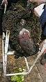 stained uterus (12 hours out)
 before re-positioning
Pigs
Rectal prolapse is a condition routinely identified in pigs on farms and at slaughterhouses. If not reduced quickly, prolapses in pigs become necrotic and infected, and risk being cannibalized by other pen mates. If the latter happens it normally results in death of the animal by septicemia, shock or faecal peritonitis.
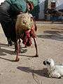
Horses and mules
Rectal prolapse occurring in horse and mule would be better termed anal prolapse, as it only involves mucous membrane moving posteriorly to form a circular protrusion outside the anus [5] The condition is not painful.
In mares after parturition, it is described as a 10 to 60 mm mucous protrusion.[6]
In young mules and foals, anal prolapse occurs with a frequency similar to that of Gasterophilus haemorrhoidalis. In extensive breeding conditions, the disease is only recognized after some days, leading to intense edema of prolapsed tissues and necrosis of the mucous membrane.
Early cases in should be treated with the application of hygroscopic substances like powdered sugar followed by purse-string suturing. When prolapsed tissues are edematous and necrotic, amputation is performed. The prognosis is fair as the removed tissues do not contain any important organs or large blood vessels.
 on a mule
 on a foal
 submucosal edema
 simple removal of prolapsed edematous tissues
References
- ↑ Page 41 in: Elizabeth D Agabegi; Agabegi, Steven S. (2008). Step-Up to Medicine (Step-Up Series). Hagerstwon, MD: Lippincott Williams & Wilkins. ISBN 0-7817-7153-6.
- ↑ Price N., Slack A., Jackson S. "Laparoscopic hysteropexy: the initial results of a uterine suspension procedure for uterovaginal prolapse." BJOG 2010;117:62–68. doi:10.1111/j.1471-0528.2009.02396. www.bjog.org
- ↑ "Vaginal rejuvenation: sounds great. What is it?". Beautycallbooks.com. Retrieved 2009-12-14.mirror
- ↑ American Urogynecologic Society (May 5, 2015), "Five Things Physicians and Patients Should Question", Choosing Wisely: an initiative of the ABIM Foundation, American Urogynecologic Society, retrieved June 1, 2015, which cites:
- Culligan, PJ (April 2012). "Nonsurgical management of pelvic organ prolapse.". Obstetrics and gynecology. 119 (4): 852–60. doi:10.1097/aog.0b013e31824c0806. PMID 22433350.
- ACOG Committee on Practice, Bulletins--Gynecology (September 2007). "ACOG Practice Bulletin No. 85: Pelvic organ prolapse.". Obstetrics and gynecology. 110 (3): 717–29. doi:10.1097/01.aog.0000263925.97887.72. PMID 17766624..
- ↑ The Merck Veterinary Manual, 3rd ed.‚ Merck and co. Inc. Rahway, N.J., USA, 1967
- ↑ V.L. Tharp, in Cattcott E.J. & Smithcors J.F. Equine Medicine and Surgery, American Veterinary Publications Inc., 2nd ed 1972, French translation Vigot Frères, Paris, France, 1974, ISBN 2-7114-0653-9. OCLC 461509749.
| Wikimedia Commons has media related to Pelvic prolapses. |