Sinus venosus
| Sinus venosus | |
|---|---|
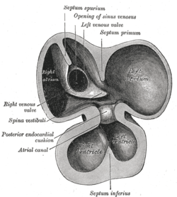 Interior of dorsal half of heart from a human embryo of about thirty days, frontal view. (Opening of sinus venosus labeled at center top.) | |
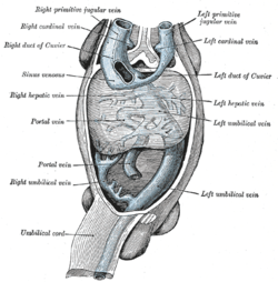 Human embryo with heart and anterior body-wall removed to show the sinus venosus and its tributaries. (Sinus venosus labeled at center left.) | |
| Details | |
| Carnegie stage | 9 |
| Identifiers | |
| Latin | sinus venosus cordis |
| Code |
TE E5.11.1.3.2.0.4; E.5.11.1.5.1.0.1 |
| TA | A12.0.00.016 |
| FMA | 70303 |
The sinus venosus is a large quadrangular cavity which precedes the atrium on the venous side of the chordate heart. In mammals, it exists distinctly only in the embryonic heart (where it is found between the two venae cavae); however, the sinus venosus persists in the adult. In the adult, it is incorporated into the wall of the right atrium to form a smooth part called the sinus venarum, also known as the venarum sinus, which is separated from the rest of the atrium by a ridge of fibres called the crista terminalis. The sinus venosus also forms the SA node and the coronary sinus.
In the embryo, the thin walls of the sinus venosus are connected below with the right ventricle, and medially with the left atrium, but are free in the rest of their extent. It receives blood from the vitelline vein, umbilical vein and common cardinal vein.
It originally starts as a paired structure but shifts towards associating only with the right atrium as the embryonic heart develops. The left portion shrinks in size and eventually forms the coronary sinus (right atrium) and oblique vein of the left atrium, whereas the right part becomes incorporated into the right atrium to form the sinus venarum.
Additional images
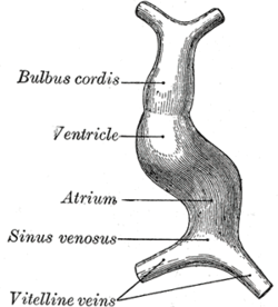 Diagram to illustrate the simple tubular condition of the heart.
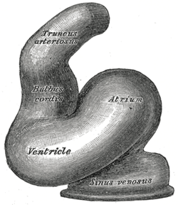 Heart of human embryo of about fourteen days.
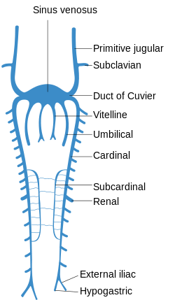 Scheme of arrangement of parietal veins.
See also
- Atrial septal defect
- Bulbus cordis
- Ducts of Cuvier
- Primitive ventricle
- Primitive atrium
- Ductus venosus
- Truncus arteriosus
- Sinus venosus atrial septal defect
External links
- 1483407420 at GPnotebook
- Anatomy photo:20:13-0102 at the SUNY Downstate Medical Center - Gross anatomy of the adult heart
- Atlas image: ht_rt_atrium at the University of Michigan Health System - "Right atrium, internal structure, anterior view"