Fetal circulation
| Fetal circulation | |
|---|---|
|
The fetal circulatory system includes three shunts to divert blood from undeveloped and partially functioning organs, as well as blood supply to and from the placenta. | |

In animals that give live birth, the fetal circulation is the circulatory system of a fetus. The term usually encompasses the entire fetoplacental circulation, which includes the umbilical cord and the blood vessels within the placenta that carry fetal blood.
The fetal (prenatal) circulation works differently from normal postnatal circulation, mainly because the lungs are not in use. Instead, the fetus obtains oxygen and nutrients from the mother through the placenta and the umbilical cord.[1] The advent of breathing and the severance of the umbilical cord prompt various neuroendocrine changes that shortly transform fetal circulation into postnatal circulation.
The fetal circulation of humans has been extensively studied by the health sciences. Much is known also of fetal circulation in other animals, especially livestock and model organisms such as mice, through the health sciences, veterinary science, and life sciences generally.
Structure
Blood from the placenta is carried to the fetus by the umbilical vein. In humans, less than a third of this enters the fetal ductus venosus and is carried to the inferior vena cava,[2] while the rest enters the liver proper from the inferior border of the liver. The branch of the umbilical vein that supplies the right lobe of the liver first joins with the portal vein. The blood then moves to the right atrium of the heart. In the fetus, there is an opening between the right and left atrium (the foramen ovale), and most of the blood flows through this hole directly into the left atrium from the right atrium, thus bypassing pulmonary circulation. The continuation of this blood flow is into the left ventricle, and from there it is pumped through the aorta into the body. Some of the blood moves from the aorta through the internal iliac arteries to the umbilical arteries, and re-enters the placenta, where carbon dioxide and other waste products from the fetus are taken up and enter the maternal circulation.[1]
Some of the blood entering the right atrium does not pass directly to the left atrium through the foramen ovale, but enters the right ventricle and is pumped into the pulmonary artery. In the fetus, there is a special connection between the pulmonary artery and the aorta, called the ductus arteriosus, which directs most of this blood away from the lungs (which are not being used for respiration at this point as the fetus is suspended in amniotic fluid).[1]
Placenta
The circulatory system of the mother is not directly connected to that of the fetus, so the placenta functions as the respiratory center for the fetus as well as a site of filtration for plasma nutrients and wastes. Water, glucose, amino acids, vitamins, and inorganic salts freely diffuse across the placenta along with oxygen. The uterine arteries carry blood to the placenta, and the blood permeates the sponge-like material there. Oxygen then diffuses from the placenta to the chorionic villus, an alveolus-like structure, where it is then carried to the umbilical vein.
After birth
At birth, when the infant breathes for the first time, there is a decrease in the resistance in the pulmonary vasculature, which causes the pressure in the left atrium to increase relative to the pressure in the right atrium. This leads to the closure of the foramen ovale, which is then referred to as the fossa ovalis. Additionally, the increase in the concentration of oxygen in the blood leads to a decrease in prostaglandins, causing closure of the ductus arteriosus. These closures prevent blood from bypassing pulmonary circulation, and therefore allow the neonate's blood to become oxygenated in the newly operational lungs.[3]
Sometimes these postnatal closures are incomplete or absent. The vessels or cross-connections remain open (patent), leading to the following conditions:
- Patent foramen ovale in the heart
- Patent ductus arteriosus in the great vessels
- Patent ductus venosus in the great vessels
Physiology
The core concept behind fetal circulation is that fetal hemoglobin (HbF)[4] has a higher affinity for oxygen than does adult hemoglobin, which allows a diffusion of oxygen from the mother's circulatory system to the fetus.
Blood pressure
It is the fetal heart and not the mother's heart that builds up the fetal blood pressure to drive its blood through the fetal circulation.
Intracardiac pressure remains identical between the right and left ventricles of the human fetus.[5]
The blood pressure in the fetal aorta is approximately 30 mmHg at 20 weeks of gestation, and increases to ca 45 mmHg at 40 weeks of gestation.[6] The fetal pulse pressure is ca 20 mmHg at 20 weeks of gestation, increasing to ca 30 mmHg at 40 weeks of gestation.[6]
The blood pressure decreases when passing through the placenta. In the arteria umbilicalis, it is ca 50 mmHg. It falls to 30 mmHg in the capillaries in the villi. Subsequently, the pressure is 20 mm Hg in the umbilical vein, returning to the heart.[7]
Flow
The blood flow through the umbilical cord is approximately 35 mL/min at 20 weeks, and 240 mL/min at 40 weeks of gestation.[8] Adapted to the weight of the fetus, this corresponds to 115 mL/min/kg at 20 weeks and 64 mL/min/kg at 40 weeks.[8] It corresponds to 17% of the combined cardiac output of the fetus at 10 weeks, and 33% at 20 weeks of gestation.[8]
Endothelin and prostanoids cause vasoconstriction in placental arteries, while nitric oxide causes vasodilation.[8] On the other hand, there is no neural vascular regulation, and catecholamines have only little effect.[8]
Prenatal heartbeat
Fetal cardiac activity (also called fetal heartbeat and usually called embryonic cardiac activity before approximately 10 weeks of gestational age) is the rate of contractions during the cardiac cycles of an embryo or fetus. The heart is not fully developed when cardiac activity becomes visible.
In cases of early pregnancy bleeding, the detection of cardiac activity is the main finding that distinguishes a viable pregnancy from a silent miscarriage.
Imaging
In the first trimester, heartbeat can be visualized and the heart rate quantified by obstetric ultrasonography. A study of 32 normal pregnancies came to the result a fetal heartbeat was visible at a mean human chorionic gonadotropin (hCG) level of 10,000 UI/l (range 8650-12,200).[9] Obstetric ultrasonography can also use doppler technique on key vessels such as the umbilical artery can detect abnormal flow.

In later stages of pregnancy, a simple Doppler fetal monitor can quantify the fetal heart rate.
During childbirth, the parameter is part of cardiotocography, which is where the fetal heartbeat and uterine contractions are continuously recorded.
Heart rates
Starting at week 5 the embryonic heart rate accelerates by 3.3 bpm per day for the next month. Before this, the embryo possesses a tubular heart.
The embryonic heart begins to beat at approximately the same rate as the mother's, which is typically 80 to 85 bpm. The approximate fetal heart rate for weeks 5 to 9 (assuming a starting rate of 80):
- Week 5 starts at 80 and ends at 103 bpm
- Week 6 starts at 103 and ends at 126 bpm
- Week 7 starts at 126 and ends at 149 bpm
- Week 8 starts at 149 and ends at 172 bpm
- At week 9 the embryonic heart tends to beat within a range of 155 to 195 bpm.
By the end of week 9, the embryonic heart has developed septa and valves, and has all four chambers.
At this point, the fetal heart rate begins to decrease, and generally falls within the range of 120 to 160 bpm by week 12.[10]

Additional images
-
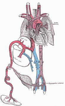
Diagram of the human feto-placental circulatory system.
-
Transvaginal ultrasonography of an embryo at 5 weeks and 5 days of gestational age with discernible heartbeat
-

Neonatal heart circulation
References
- 1 2 3 Whitaker, Kent (2001). "Fetal Circulation". Comprehensive Perinatal and Pediatric Respiratory Care. Delmar Thomson Learning. pp. 18–20. ISBN 978-0-7668-1373-1.
- ↑ Kiserud, T.; Rasmussen, S.; Skulstad, S. (2000). "Blood flow and the degree of shunting through the ductus venosus in the human fetus". American Journal of Obstetrics and Gynecology. 182 (1 Pt 1): 147–153. doi:10.1016/S0002-9378(00)70504-7. PMID 10649170.
- ↑ Le, Tao; Bhushan, Vikas; Vasan, Neil (2010). First Aid for the USMLE Step 1: 2010 20th Anniversary Edition. USA: McGraw-Hill. p. 123. ISBN 978-0-07-163340-6.
- ↑ Edoh D, Antwi-Bosaiko C, Amuzu D (March 2006). "Fetal hemoglobin during infancy and in sickle cell adults". African Health Sciences. 6 (1): 51–54. doi:10.5555/afhs.2006.6.1.51. PMC 1831961
 . PMID 16615829.
 - ↑ Johnson P, Maxwell DJ, Tynan MJ, Allan LD (2000). "Intracardiac pressures in the human fetus". Heart. 84 (1): 59–63. doi:10.1136/heart.84.1.59. ISSN 0007-0769.
- 1 2 Struijk, P. C.; Mathews, V. J.; Loupas, T.; Stewart, P. A.; Clark, E. B.; Steegers, E. A. P.; Wladimiroff, J. W. (2008). "Blood pressure estimation in the human fetal descending aorta". Ultrasound in Obstetrics and Gynecology. 32 (5): 673–81. doi:10.1002/uog.6137. PMID 18816497.
- ↑ "Fetal and maternal blood circulation systems". Swiss Virtual Campus. Retrieved June 29, 2011.
- 1 2 3 4 5 Kiserud, Torvid; Acharya, Ganesh (2004). "The fetal circulation". Prenatal Diagnosis. 24 (13): 1049–59. doi:10.1002/pd.1062. PMID 15614842.
- ↑ Giacomello F, Magliocchetti P, Loyola G, Giovarruscio M (1993). "[Serum beta hCG levels and transvaginal echography in the early phases of pregnancy]". Minerva Ginecol (in Italian). 45 (7-8): 333–7. PMID 8414139.
- ↑ FetalSure. Fetal Heart and Heartbeat Facts. Available at http://www.fetalsure.com/fetal-heart.html. Retrieved 9 August 2010.