Sessile serrated adenoma
| Sessile serrated adenoma | |
|---|---|
 | |
| Micrograph of a sessile serrated adenoma. H&E stain. | |
| Classification and external resources |
In gastroenterology, a sessile serrated adenoma (abbreviated SSA), also known as sessile serrated polyp (abbreviated SSP), is a premalignant flat (or sessile) lesion of the colon, predominantly seen in the cecum and ascending colon.
SSAs are thought to lead to colorectal cancer through the (alternate) serrated pathway.[1][2] This differs from most colorectal cancer, which arises from mutations starting with inactivation of the APC gene.
Multiple SSAs may be part of the serrated polyposis syndrome.[3]
Symptoms
SSAs, generally, are asymptomatic. They are typically identified on a colonoscopy and excised for a definitive diagnosis and treatment.
Diagnosis
SSAs are diagnosed by their microscopic appearance; histomorphologically, they are characterized by (1) basal dilation of the crypts, (2) basal crypt serration, (3) crypts that run horizontal to the basement membrane (horizontal crypts), and (4) crypt branching. The most common of these features is basal dilation of the crypts.
Unlike traditional colonic adenomas (e.g. tubular adenoma, villous adenoma), they do not (typically) have nuclear changes (nuclear hyperchromatism, nuclear crowding, elliptical/cigar-shaped nuclei).
Serrated polyposis syndrome
The serrated polyposis syndrome (SPS) is a relatively rare condition characterized by multiple and/or large serrated polyps of the colon. Diagnosis of this disease is made by the fulfillment of any of the World Health Organization’s (WHO) clinical criteria.[4]
Treatment
Complete removal of a SSA is considered curative.
Several SSAs confer a higher risk of subsequently finding colorectal cancer and warrant more frequent surveillance. The surveillance guidelines are the same as for other colonic adenomas. The surveillance interval is dependent on (1) the number of adenomas, (2) the size of the adenomas, and (3) the presence of high-grade microscopic features.[5]
Additional images
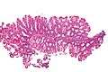 Low magnification micrograph of a SSA.
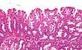 Intermediate magnification micrograph of a SSA.
 High magnification micrograph of a SSA showing crypt branching.
See also
References
- ↑ Rüschoff J, Aust D, Hartmann A (2007). "[Colorectal serrated adenoma: diagnostic criteria and clinical implications]". Verh Dtsch Ges Pathol (in German). 91: 119–25. PMID 18314605.
- ↑ Mäkinen MJ (January 2007). "Colorectal serrated adenocarcinoma". Histopathology. 50 (1): 131–50. doi:10.1111/j.1365-2559.2006.02548.x. PMID 17204027.
- ↑ Rosty, C.; Parry, S.; Young, JP. (2011). "Serrated polyposis: an enigmatic model of colorectal cancer predisposition.". Patholog Res Int. 2011: 157073. doi:10.4061/2011/157073. PMC 3109311
 . PMID 21660283.
 - ↑ World J Gastroenterol 2012 May 28; 18(20): 2452-2461
- ↑ Levine JS, Ahnen DJ (December 2006). "Clinical practice. Adenomatous polyps of the colon" (PDF). N. Engl. J. Med. 355 (24): 2551–7. doi:10.1056/NEJMcp063038. PMID 17167138.
External links
- Intestinal adenomas - halflytely.com.
- Pathology of Serrated Colon Adenomas - Medscape