Radiographic equipment
This is a page devoted to the basic equipment used for radiographic work, both medical and industrial.
Photon sources
There are many types of sources for high energy X-ray and gamma photons.[1]
X-ray sources
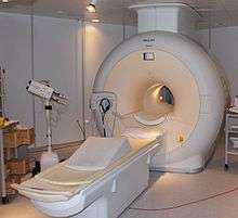
X-ray generators are made of a high voltage power supply that is applied on a usually sealed X-ray tube. This produces the emission of electrons from the cathode of the tube and the emission of X-rays when these hit a target located at the anode side.
In industrial radiography, energy goes from 20 to several hundreds of kV according to the application. In medical radiography voltage from 20 kV in mammography up to 150 kV for chest radiography are used for diagnostic. Energy can go up to 250 kV for radiotherapy applications.
Radioisotope sources
These have the advantage that they do not need a supply of electrical power to function, but they have the disadvantage that they can not be turned off. Also it is difficult, using radioactivity, to create a small and compact source that offers the photon flux possible with a normal sealed X-ray tube.
It might be possible to use Cs-137 as a photon source for radiography but this isotope has the disadvantage that it is always diluted with inactive caesium isotopes. This means that it is difficult to get a physically small source, a large radioactive volume of the source will make it impossible to get the finest detail from a radiographic examination.
Both cobalt-60 and caesium-137 have only a few gamma energies, which makes them close to monochromatic. The photon energy of cobalt-60 is higher than that of caesium-137, which allows cobalt sources to be used to examine thicker sections of metals than those that could be examined with Cs-137. Iridium-192 has a lower photon energy than cobalt-60 and its gamma spectrum is complex (many lines of very different energies), but this can be an advantage as this can give better contrast for the final photographs.
It has been known for many years that an inactive iridium or cobalt metal object can be machined to size. In the case of cobalt it is common to alloy it with nickel to improve the mechanical properties. In the case of iridium a thin wire or rod could be used. These precursor materials can then be placed within stainless steel containers, which are leak tested before being converted into radioactive sources. These objects can be processed by neutron activation to form gamma emitting radioisotopes. The stainless steel has only a small ability to be activated and the small activity due to 55Fe and 63Ni are unlikely to pose a problem in the final application because these isotopes are beta emitters, which have very weak gamma emission. The 59Fe that might form has a short half-life, so by allowing a cobalt source to stand for a year much of this isotope decays away.
The source is often a small object that must be transported to the work site in a shielded container. It is normal to position the film, clear the area where the work is to be done, and add shielding (collimators) to reduce the size of the controlled area before exposing the radioactive source. A series of different designs have been developed for radiographic "cameras". Rather than the "camera" being a device that accepts photons to record a picture, the "camera" in industrial radiography is the radioactive photon source.
Torch design of radiographic cameras
One design is best thought of as being like a torch. The radioactive source is placed inside a shielded box, a hinge allowed part of the shielding to be peeled back exposing the source so allowing the photons to leave the radiography camera.

Red represents the radioactive source; blue/green, the shielding; and yellow, the gamma rays
Another design for a torch is one where the source is placed in a metal wheel, this can turn inside the camera to move between the exposed and storage sites.

The radioactive source is red and the gamma rays are yellow
.
Cable-based design of radiographic cameras
One group of designs uses a radioactive source that comes out on a cable from a shielded container . One such unit was involved in an accident in Bolivia. This method is similar to brachytherapy when performed with the remote afterloading method. An example of a cable-based design would have the source stored in a block of lead or depleted uranium with an S-shaped passage through the block. In its safe position the source is held in the centre of the block by a metal wire extending in both directions. To use the source, a drive cable is attached to one end of the wire, and a guide tube is attached to the opposite side of the block. A hand-operated winch pushes the source out of the shield and along the guide tube to where it is needed.

Microsecond X-ray pulses
It is possible using a particle accelerator to generate a short pulse of high energy electrons, these electrons are used to create X-rays by braking radiation.. The X-rays are detected using a semiconductor detector, which is an array of silicon diodes. Such equipment has been used for the X-ray version of high speed flash photography. For example diesel fuel that has been doped with cerium has been used to investigate the operation of fuel injectors in a diesel engine..
Some examples of radiography using a 5 MeV electron LINAC driving a bremsstrahlung source (1 mm Tungsten on a 9 mm copper sheet) can be seen here.
As an alternative high energy pulsed proton beams can be used for the high speed examination of objects.
Neutron sources
In some cases, industrial radiography is done with neutrons. This type of radiography is called Neutron Radiography (NR, Nray, N-Ray) or Neutron Imaging. Neutron Radiography can see very different things than X-rays, because neutrons can pass with ease through lead and steel but are stopped by plastics, water and oils. Neutron sources include radioactive (241Am/Be and Cf) sources, electrically driven D-T reactions in vacuum tubes and conventional critical nuclear reactors. It might be possible to use a neutron amplifier to increase the neutron flux.