Medical radiography
| Medical radiography | |
|---|---|
| Diagnostics | |
| ICD-10-PCS | B?0, B?1, B?2 |
| ICD-9-CM | 87, 88.0-88.6 |
| MeSH | D011859 |
| OPS-301 code | 3-10...3-13, 3-20...3-26 |
Radiography is the use of ionizing electromagnetic radiation such as X-rays to view objects. Although not technically radiographic techniques, imaging modalities such as PET and MRI are sometimes grouped in radiography because the radiology department of hospitals handle all forms of imaging. Treatment using radiation is known as radiotherapy.
History
Radiography started in 1895 with the discovery of X-rays (later also called Röntgen rays after the man who first described their properties in rigorous detail), a type of electromagnetic radiation. Soon these found various applications, from helping to find shoes that fit, to the more lasting medical uses. X-rays were put to diagnostic use very early, before the dangers of ionising radiation were discovered. Initially, many groups of staff conducted radiography in hospitals, including physicists, photographers, doctors, nurses, and engineers. The medical speciality of radiology grew up around the new technology, and this lasted many years. When new diagnostic tests involving X-rays were developed, it was natural for the radiographers to be trained and adopt this new technology. This happened first with fluoroscopy, computed tomography (1960s), and mammography. Ultrasound (1970s) and magnetic resonance imaging (1980s) was added to the list of skills used by radiographers because they are also medical imaging, but these disciplines do not use ionising radiation or X-rays. Although a nonspecialist dictionary might define radiography quite narrowly as "taking X-ray images", this has only been part of the work of radiographers, and radiologists for a very long time. X-rays are also exploited by industrial radiographers in the field of nondestructive testing, where the newer technology of ultrasound is also used.
Diagnostic radiography
Diagnostic radiography involves the use of both ionising radiation and non-ionising radiation to create images for medical diagnoses. The predominant test is still the X-ray (the word X-ray is often used for both the test and the actual film or digital image). X-rays are the second most commonly used medical tests, after laboratory tests. This application is known as diagnostic radiography. Since the body is made up of various substances with differing densities, X-rays can be used to reveal the internal structure of the body on film by highlighting these differences using attenuation, or the absorption of X-ray photons by the denser substances (like calcium-rich bones). Anatomy discipline which involves the study of anatomy through the use of radiographic films is known as radiographic anatomy.
Medical diagnostic radiography is undertaken by a specially trained professional called radiographers or radiologic technologists.
There are several sub-specialities:
Projection radiography
For the main article see Projectional Radiography
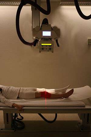
.jpg)
The creation of images by exposing an object to X-rays or other high-energy forms of electromagnetic radiation and capturing the resulting remnant beam (or "shadow") as a latent image is known as "projection radiography." The "shadow" may be converted to light using a fluorescent screen, which is then captured on photographic film, it may be captured by a phosphor screen to be "read" later by a laser (CR), or it may directly activate a matrix of solid-state detectors (DR—similar to a very large version of a CCD in a digital camera). Bone and some organs (such as lungs) especially lend themselves to projection radiography. It is a relatively low-cost investigation with a high diagnostic yield. The difference between soft and hard body parts stems mostly from the fact that carbon has a very low X-ray cross section compared to calcium.
Projection radiography uses X-rays in different amounts and strengths depending on what body part is being imaged:
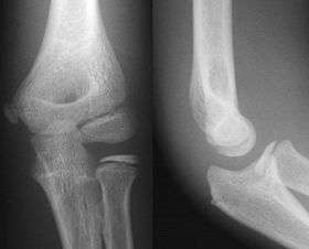
- Hard tissues such as bone require a relatively high energy photon source, and typically a tungsten anode is used with a high voltage (50-150 kVp) on a 3-phase or high-frequency machine to generate bremsstrahlung or braking radiation. Bony tissue and metals are denser than the surrounding tissue, and thus by absorbing more of the X-ray photons they prevent the film from getting exposed as much.[1] Wherever dense tissue absorbs or stops the X-rays, the resulting X-ray film is unexposed, and appears translucent blue, whereas the black parts of the film represent lower-density tissues such as fat, skin, and internal organs, which could not stop the X-rays. This is usually used to see bony fractures, foreign objects (such as ingested coins), and used for finding bony pathology such as osteoarthritis, infection (osteomyelitis), cancer (osteosarcoma), as well as growth studies (leg length, achondroplasia, scoliosis, etc.).
- Soft tissues are seen with the same machine as for hard tissues, but a "softer" or less-penetrating X-ray beam is used. Tissues commonly imaged include the lungs and heart shadow in a chest X-ray, the air pattern of the bowel in abdominal X-rays, the soft tissues of the neck, the orbits by a skull X-ray before an MRI to check for radiopaque foreign bodies (especially metal), and of course the soft tissue shadows in X-rays of bony injuries are looked at by the radiologist for signs of hidden trauma (for example, the famous "fat pad" sign on a fractured elbow).
- Dental radiography uses a small radiation dose with high penetration to view teeth, which are relatively dense. A dentist may examine a painful tooth and gum using X-ray equipment. The machines used are typically single-phase pulsating DC, the oldest and simplest sort. Dental technicians or the dentist may run these machines; radiographers are not required by law to be present.
- Mammography is an X-ray examination of breasts and other soft tissues. This has been used mostly on women to screen for breast cancer, but is also used to view male breasts, and used in conjunction with a radiologist or a surgeon to localise suspicious tissues before a biopsy or a lumpectomy. Breast implants designed to enlarge the breasts reduce the viewing ability of mammography, and require more time for imaging as more views need to be taken. This is because the material used in the implant is very dense compared to breast tissue, and looks white (clear) on the film. The radiation used for mammography tends to be softer (has a lower photon energy) than that used for the harder tissues. Often a tube with a molybdenum anode is used with about 30 000 volts (30 kV), giving a range of X-ray energies of about 15-30 keV. Many of these photons are "characteristic radiation" of a specific energy determined by the atomic structure of the target material (Mo-K radiation).
Other modalities are used in radiography when traditional projection X-ray cannot image what doctors want to see. Below are other modalities included within radiography; they are only summaries and more specific information can be viewed by going to their individual pages:
Fluoroscopy
Fluoroscopy is a term invented by Thomas Edison during his early X-ray studies. The name refers to the fluorescence he saw while looking at a glowing plate bombarded with X-rays.
This is a technique that provides moving projection radiographs of lower quality. Fluoroscopy is mainly performed to view movement (of tissue or a contrast agent), or to guide a medical intervention, such as angioplasty, pacemaker insertion, or joint repair/replacement. The latter are often carried out in the operating theatre, using a portable fluoroscopy machine called a C-arm. It can move around the surgery table and make digital images for the surgeon.
Biplanar Fluoroscopy
Biplanar Fluoroscopy works the same as single plane fluoroscopy except displaying two planes at same the time. This gives the advantage to work in two planes which is necessary for orthopedic and spinal surgery. The operation time is greatly reduced since no re-positioning is required.
Angiography
Angiography is the use of fluoroscopy to view the cardiovascular system. An iodine-based contrast is injected into the bloodstream and watched as it travels around. Since liquid blood and the vessels are not very dense, a contrast with high density (like the large iodine atoms) is used to view the vessels under X-ray. Angiography is used to find aneurysms, leaks, blockages (thromboses), new vessel growth, and placement of catheters and stents. Balloon angioplasty is often done with angiography.
Dual energy X-ray absorptiometry
DEXA, or bone densitometry, is used primarily for osteoporosis tests. It is not projection radiography, as the X-rays are emitted in 2 narrow beams that are scanned across the patient, 90 degrees from each other. Usually the hip (head of the femur), lower back (lumbar spine) or heel (calcaneum) are imaged, and the bone density (amount of calcium) is determined and given a number (a T-score). It is not used for bone imaging, as the image quality is not good enough to make an accurate diagnostic image for fractures, inflammation etc. It can also be used to measure total body fat, though this isn't common. The radiation dose received from DEXA scans is very low, much lower than projection radiography examinations.
Computed tomography
Computed tomography or CT scan (previously known as CAT scan, the "A" standing for "axial") uses a high amount of ionizing radiation (in the form of X-rays) in conjunction with a computer to create images of both soft and hard tissues. These images look as though the patient was sliced like bread (thus, "tomography"-- "tomo" means "slice"). The machine looks similar to an MRI machine to many patients, but is not related. The exams are generally short, most lasting only as long as a breath-hold. Contrast agents are often used, depending on the tissues needing to be seen. Radiographers perform these examinations, sometimes in conjunction with a radiologist (for instance, when a radiologist performs a CT-guided biopsy).
Technical considerations
X-ray photons are formed in events involving electrons and are the mainly form of ionizing electromagnetic radiation used in medical radiography. This radiation is much more energetic than the more familiar types such as radio waves and visible light. Proper production and detection of photons are important in the creation of good radiograms.
Photon production
X-ray radiation for medical imaging is typically produced by X-ray tubes, which operate through bombarding the anode with high energy electrons emitted from a hot cathode(the stream of electron with high speed interact to the target 'anode' from cathode due to this the generation of x-ray occur.). Image sharpness, contrast, and patient dosage are important considerations in medical radiography and these requirements determined the desired energies of the tube, the type of material used on the anode, and the method in which the power is generated to drive the tube. Although the technical definition of x-rays range from 1-700 keV, medical x-rays typically use 5-150 keV x-rays. The photons emitted come in discrete bands of energy corresponding to the material of the anode, and the undesired bands are removed. Choice of the anode and its emitted radiation energies depends on the application and the tissues being imaged, for instance molybdenum is often used in mammography because of its 20 keV x-rays. Too high radiation energies will result in poor pictures since the radiation cannot be readily attenuated, however too low energies will increase the radiation dosage of the patient without improvements in image quality.
Sharpness of a radiographic image is strongly determined by the size of the x-ray source. This is determined by the area of the electron beam hitting the anode. A large photon source results in more blurring in the final image and is worsened by an increase in image formation distance. This blurring can be measured as a contribution to the modulation transfer function of the imaging system.
Power generation
The power used by the x-ray tube is generated by a specialized generator, which supplies the voltage and current required to drive the tube. The generator needs to supply high voltages with small exposure times. An exposure thus can be described by two factors:
- The peak voltage of the cathode to anode
- The milliampere seconds exposure time
These variables can be controlled by the operator but are more typically assigned automatically by the x-ray machinery through sampling the emitted radiation. Power generators convert standard 120 or 220 volt AC to higher DC voltages and typically employ rectified and filtered multiphase transformers which maintain a constant voltage and can be turn rapidly on and off for millisecond exposures.
Photon detection
Photons images that have been shadowed from an imaging subject must be detected at high fidelity and resolution to allow for diagnosis. There are three main types of image detection methods used namely: film/screens, image intensifiers, and digital detectors, with the latter fast becoming the standard for x-ray image detection. The ability of an x-ray detector to produce high-quality images is determined largely by the modulation transfer function (MTF) and detective quantum efficiency (DQE) of the system.
A Bucky-Potter grid may be placed between the patient and the detector to reduce the quantity of scattered x-rays that reach the detector. This improves the contrast resolution of the image, but also increases radiation exposure for the patient.
Film/Screens
X-ray film is almost always used in conjunction with an x-ray sensitive screen, because high resolution film is quite poor at detecting x-rays. These screens contain rare earth minerals and phosphor materials which convert x-ray radiation into visible light (of lower electromagnetic energy) to which the film is more sensitive. Screens generally have to have high resolution, good contrast, and a wide dynamic range, the last two being antithetic properties. The sensitivity of the screen inversely affects its resolution, since more sensitive screens are generally thicker causing more blurring as emitted light spreads out.
The film's speed also plays a factor in image quality. Higher speeds are more sensitive to photons but are generally lower in resolution and more susceptible to noise. Lower speed films produce images of good resolution and dynamic range but require more photons for exposure and increase the radiation dosage to the subject.
Image intensifiers and array detectors
Image intensifiers are analog devices that readily convert the acquired x-ray image into one visible on a video screen. This device is made of a vacuum tube with a wide input surface coated on the inside with caesium iodide (CsI). When hit by x-rays material phosphors which causes the photocathode adjacent to it to emit electrons. These electron are then focus using electron lenses inside the intensifier to an output screen coated with phosphorescent materials. The image from the output can then be recorded via a camera and displayed.
Digital devices known as array detectors are becoming more common in fluoroscopy. These devices are made of discrete pixelated detectors known as TFTs which can either work indirectly by using photo detectors that detect light emitted from a scintillator material such as CsI, or directly by capturing the electrons produced when the x-rays hit the detector. Direct detector do not tend to experience the blurring or spreading effect caused by phosphorescent scintillators of or film screens since the detectors are activated directly by x-ray photons.
Obsolete terminology
The term skiagrapher was used until about 1918 to mean radiographer.
The word was derived from Ancient Greek words for 'shadow' and 'writer'.[2]
See also
References
- ↑ Advances in kilovoltage x-ray beam dosimetry, http://iopscience.iop.org/0031-9155/59/6/R183/article
- ↑ The Electrical world. Electrical World. 1896. pp. 372–. Retrieved 27 June 2011.
- Shroy, Jr., Robert E. (1995). "X-Ray equipment". In Bronzino, J.D. The Biomedical Engineering handbook. CRC Press and IEEE Press. pp. 953–960. ISBN 0-8493-8346-3.
- Bushberg, Jerrold T.; Seibert, J. Anthony; Leidholdt Jr., Edwin M.; Boone, John M. (2001). The Essential Physics of Medical Imaging (2nd ed.). Lippincott Williams & Wilkins. ISBN 978-0-683-30118-2.
- Herman, Gabor T. (2009). Fundamentals of Computerized Tomography: Image Reconstruction from Projections (2nd ed.). Springer. ISBN 978-1-85233-617-2.
- Kodak. (http://www.kodak.com/global/en/health/productsByType/index.jhtml?pq-path=2/521/2970)
- Agfa. (http://www.piribo.com/publications/medical_devices/mdc/agfa_medical.html)
- Yu, Shi-Bao; Alan D. Watson (1999). "A review on the subject of medical X-ray examinations and metal based contrast agents". Chemical Reviews. 99 (9): 2353–2378. doi:10.1021/cr980441p. PMID 11749484.
- Baker, Alan; Stuart Dutton (Ed.) (2004). Composite Materials for Aircraft Structures. American Institute of Aeronautics & Ast. ISBN 1-56347-540-5.
External links
 Media related to Medical X-rays at Wikimedia Commons
- NIST's XAAMDI: X-Ray Attenuation and Absorption for Materials of Dosimetric Interest Database
- NIST's XCOM: Photon Cross Sections Database
- NIST's FAST: Attenuation and Scattering Tables
- Major John Hall-Edwards, British radiography pioneer
- A lost industrial radiography source event
- UN information on the security of industrial sources
- RadiologyInfo - The radiology information resource for patients: Radiography (X-rays)
- Nick Oldnall's radiography site
- MedPix Free Medical Image Database (radiology website)