Liver tumor
| Liver tumor | |
|---|---|
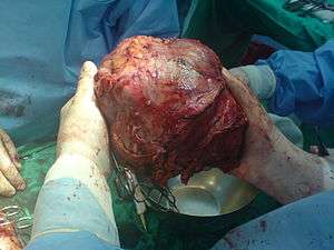 | |
| left lobe liver tumor in 50-year-old man operated in King Saud Medical Complex, Riyadh, Saudi Arabia | |
| Classification and external resources | |
| MeSH | D008113 |
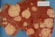
Liver tumors or hepatic tumors are tumors or growths on or in the liver (medical terms pertaining to the liver often start in hepato- or hepatic from the Greek word for liver, hepar). Several distinct types of tumors can develop in the liver because the liver is made up of various cell types. These growths can be benign or malignant (cancerous). They may be discovered on medical imaging (even for a different reason than the cancer itself), or may be present in patients as an abdominal mass, hepatomegaly, abdominal pain, jaundice, or some other liver dysfunction.
Classification
There are many forms of liver tumors:
Malignant (cancerous)

- Most cases are metastases from other tumors, frequently of the GI tract (like colon cancer, carcinoid tumors mainly of the appendix, etc.), but also from breast cancer, ovarian cancer, lung cancer, renal cancer, prostate cancer, etc.
- The most frequent, malignant, primary liver cancer is hepatocellular carcinoma (also named hepatoma, which is a misnomer because adenomas are usually benign).
- More rare primary forms of liver cancer include cholangiocarcinoma, mixed tumors, sarcoma and hepatoblastoma; a rare malignant tumor in children.
Benign

There are several types of benign liver tumor.
Hemangiomas: These are the most common type of benign liver tumor, found in up to 7% of autopsy specimens. They start in blood vessels. Most of these tumors do not cause symptoms and do not need treatment. Some may bleed and need to be removed if it is mild to severe. A rare tumor is Infantile hemangioendothelioma.
Hepatic adenomas: These benign epithelial liver tumors develop in the liver and are also an uncommon occurrence, found mainly in women using estrogens as contraceptives, or in cases of steroid abuse. They are, in most cases, located in the right hepatic lobe and are frequently seen as solitary. The size of adenomas range from 1 to 30 cm. Symptoms associated with hepatic adenomas are all associate with large lesions which can cause intense abdominal pain. Over the last few decades there has been an increase with occurrences of this specific type of adenoma. The prognosis for these tumors has still not been mastered. Some correlations have been made such as malignant transformation, spontaneous hemorrhage, and rupture.
Focal nodular hyperplasia (FNH) is the second most common tumor of the liver. This tumor is the result of a congenital arteriovenous malformation hepatocyte response. This process is one in which all normal constituents of the liver are present, but the pattern by which they are presented is abnormal. Even though those conditions exist the liver still seems to perform in the normal range. Other types include nodular regenerative hyperplasia and hamartoma.
References
External links
| Wikimedia Commons has media related to Ultrasound images of liver tumors. |
- Radiology of Hemangioma at USUHS - MedPix
- The Liver Cancer Web Page at Johns Hopkins University
- Liver cancer at Mayo Clinic