Cathepsin S


| View/Edit Human | View/Edit Mouse |
Cathepsin S, also known as CTSS, is a protein that in humans is encoded by the CTSS gene.[4] Transcript variants utilizing alternative polyadenylation signals exist for this gene.[4]
The protein encoded by this gene, a member of the peptidase C1 family, is a lysosomal cysteine protease that may participate in the degradation of antigenic proteins to peptides for presentation on MHC class II molecules. The encoded protein can function as an elastase over a broad pH range in alveolar macrophages. Transcript variants utilizing alternative polyadenylation signals exist for this gene.[4]
Function
Cathepsin S is a lysosomal enzyme that belongs to the papain family of cysteine proteases. While a role in antigen presentation has long been recognized, it is now understood that cathepsin S has a role in itch and pain, or nociception . The nociceptive activity results from cathepsin S functioning as a signaling molecule via activation of protease-activated receptors 2 and 4 members of the G-protein coupled receptor family.[5]
Cathepsin S is expressed by antigen presenting cells including macrophages, B-lymphocytes, dendritic cells and microglia. Cathepsin S is expressed by some epithelial cells. Its expression is markedly increased in human keratinocytes following stimulation with interferon-gamma and its expression is elevated in psoriatic keratinocytes due to stimulation by proinflammatory factors. In contrast, cortical thymic epithelial cells do not express cathepsin S.
While pH optima of many lysosomal proteases are acidic, cathepsin S is an exception. This enzyme remains catalytically active under the neutral pH and has pH optimum between the pH values 6.0 and 7.5. Many lysosomal proteases are trapped inside the lysosome due to a problem with their stability. In contrast, cathepsin S remains stable and has a physiological role outside the lysosome. Immune cells, including macrophages and microglia, secrete cathepsin S in response to inflammatory mediators including lipopolysaccharides, proinflammatory cytokines and neutrophils. In vitro, cathepsin S retains some enzyme activity in the presence of 3M urea. Cathepsin S is produced as a zymogen and is activated by processing.
The activity of cathepsin S is tightly regulated by its endogenous inhibitor, cystatin C, which also has a role in antigen presentation. Cystatin A and B have a lower activity compared to cystatin C.
The active cleavage sites -(-Val-Val-Arg-)- of cathepsin S are supposed to have at least two amino acids surrounding it from each side.
While lysosomal proteases terminally degrade proteins in lysosomes, cathepsin S has own distinctive physiological role.
Role in antigen presentation
This enzyme has a critical role in antigen presentation. Major histocompatibility complex class II molecules interact with small peptide fragments for presentation on the surface of antigen-presenting immune cells. Cathepsin S participates in the degradation of the invariant or Ii chain that prevents loading the antigen into the complex. This degradation occurs in the lysosome. Chronologically, action of cathepsin S follows two cleavages performed by aspartyl proteases. Cathepsin S cleaves the remaining fragment of Ii (IiP1) and leaves a small part of Ii known as CLIP which stays directly associated with the complex.
Proteolytic degradation of Ii is important since it facilitates dissociation of CLIP from MHC II and then, complex can load the selected antigen. After loading the antigen, MHC II molecule moves to the cell surface. Thus, we can speculate that overexpression of cathepsin S may lead to premature degradation of Ii, occasional loading of MHC II and an autoimmune attack. Contrary, inhibition of cathepsin S will lead to a delay in degradation of Ii and loading the antigen into MHC II as well as inappropriate presence of uncleaved Li-fragments in MHC II on the cell surface. It will impair and weaken the immune response. For instance, this kind of MHC II will not be very efficient to induce proliferation of T-cells.
In macrophages, cathepsin S can be replaced by cathepsin F.
Role in degradation of ECM
Secreted cathepsin S cleaves some extracellular matrix (ECM) proteins. Cathepsin S may be considered the most potent elastase known. The list of proposed cathepsin S substrates includes laminin, fibronectin elastin, osteocalcin and some collagens. It also cleaves chondroitin sulfate, heparan sulfate and proteoglycans of the basal membrane. Cathepsin S plays an active role in blood vessels permeability and angiogenesis due to its elastolytic and collagenolytic activities. For instance, cleavage of laminin-5 by cathepsin S leads to generation of proangiogenic peptides. The expression of cathepsin S can be triggered by proinflammatory factors secreted by tumor cells.
In tumorogenesis, cathepsin S promotes a tumor growth.
Nociception
Cathepsin S has a role in nociception, including itch and gastrointestinal pain. The mechanism by which cathepsin S leads to itch and pain is consistent with the capacity of this cysteine protease to activate protease-activated receptors 2 and 4.[6][7]
Cathepsin S inhibitors
Synthetic inhibitors of cathepsin S participated in numerous preclinical studies for the immune disorders including rheumatoid arthritis. Currently, at least one of them participates in a clinical trial for psoriasis. LHVS (morpholinurea-leucine-homophenylalanine-vinylsulfone-phenyl) is the most extensively studied synthetic inhibitor of cathepsin S. IC50 of LHVS is about 5 nM. Inhibition of cathepsin S by LHVS has shown to be neuroprotective after traumatic brain injury.[8] The list of commercial inhibitors also includes paecilopeptin (acetyl-Leu-Val-CHO) and some others.
Clinical significance
Cathepsin S has been shown to be a significant prognostic factor for patients with type IV astrocytomas (glioblastoma multiforme), and its inhibition has shown improvement in survival time by mean average 5 months. This is because the cysteine enzyme can no longer act together with other proteases to break up the brain extracellular matrix. So the spread of the tumor is halted. Scientists have just announced that this enzyme predicts death, as it has been shown to be associated with both heart disease and cancer.
See also
References
- ↑ "Drugs that physically interact with Cathepsin S view/edit references on wikidata".
- ↑ "Human PubMed Reference:".
- ↑ "Mouse PubMed Reference:".
- 1 2 3 "Entrez Gene: CTSS cathepsin S".
- ↑ Reddy, VB; Sun, S; Azimi, E; Elmariah, SB; Dong, X; Lerner, EA (28 July 2015). "Redefining the concept of protease-activated receptors: cathepsin S evokes itch via activation of Mrgprs.". Nature Communications. 6: 7864. doi:10.1038/ncomms8864. PMC 4520244
 . PMID 26216096.
 - ↑ Elmariah SB, Reddy VB, Lerner EA (June 25, 2014). "Cathepsin S signals via PAR2 and generates a novel tethered ligand receptor agonist.". PLOS ONE. 9 (6): e99702. doi:10.1371/journal.pone.0099702. PMC 4070910. PMID 24964046.
- ↑ Reddy VB, Sun S, Azimi E, Elmariah SB, Dong X, Lerner EA (28 July 2015). "Redefining the concept of protease-activated receptors: cathepsin S evokes itch via activation of Mrgprs". Nature Communications. 6: 7864. doi:10.1038/ncomms8864. PMC 4520244. PMID 26216096.
- ↑ "Inhibition of cathepsin S produces neuroprotective effects after traumatic brain injury in mice.". Mediators Inflamm. 2013 (2013): 187873. Oct 24, 2013. doi:10.1155/2013/187873. PMID 24282339.
Further reading
- Shi GP, Munger JS, Meara JP, et al. (1992). "Molecular cloning and expression of human alveolar macrophage cathepsin S, an elastinolytic cysteine protease.". J. Biol. Chem. 267 (11): 7258–62. PMID 1373132.
- Wiederanders B, Brömme D, Kirschke H, et al. (1992). "Phylogenetic conservation of cysteine proteinases. Cloning and expression of a cDNA coding for human cathepsin S.". J. Biol. Chem. 267 (19): 13708–13. PMID 1377692.
- Ritonja A, Colić A, Dolenc I, et al. (1991). "The complete amino acid sequence of bovine cathepsin S and a partial sequence of bovine cathepsin L.". FEBS Lett. 283 (2): 329–31. doi:10.1016/0014-5793(91)80620-I. PMID 2044774.
- Munger JS, Haass C, Lemere CA, et al. (1995). "Lysosomal processing of amyloid precursor protein to A beta peptides: a distinct role for cathepsin S.". Biochem. J. 311 (1): 299–305. PMC 1136152. PMID 7575468.
- Lemere CA, Munger JS, Shi GP, et al. (1995). "The lysosomal cysteine protease, cathepsin S, is increased in Alzheimer's disease and Down syndrome brain. An immunocytochemical study.". Am. J. Pathol. 146 (4): 848–60. PMC 1869262. PMID 7717452.
- Hall A, Håkansson K, Mason RW, et al. (1995). "Structural basis for the biological specificity of cystatin C. Identification of leucine 9 in the N-terminal binding region as a selectivity-conferring residue in the inhibition of mammalian cysteine peptidases.". J. Biol. Chem. 270 (10): 5115–21. doi:10.1074/jbc.270.10.5115. PMID 7890620.
- Balbín M, Hall A, Grubb A, et al. (1994). "Structural and functional characterization of two allelic variants of human cystatin D sharing a characteristic inhibition spectrum against mammalian cysteine proteinases.". J. Biol. Chem. 269 (37): 23156–62. PMID 8083219.
- Shi GP, Webb AC, Foster KE, et al. (1994). "Human cathepsin S: chromosomal localization, gene structure, and tissue distribution.". J. Biol. Chem. 269 (15): 11530–6. PMID 8157683.
- Turk B, Stoka V, Turk V, et al. (1996). "High-molecular-weight kininogen binds two molecules of cysteine proteinases with different rate constants.". FEBS Lett. 391 (1–2): 109–12. doi:10.1016/0014-5793(96)00611-4. PMID 8706894.
- Baumgrass R, Williamson MK, Price PA (1997). "Identification of peptide fragments generated by digestion of bovine and human osteocalcin with the lysosomal proteinases cathepsin B, D, L, H, and S". J. Bone Miner. Res. 12 (3): 447–55. doi:10.1359/jbmr.1997.12.3.447. PMID 9076588.
- Würl P, Taubert H, Meye A, et al. (1997). "Immunohistochemical and clinical evaluation of cathepsin expression in soft tissue sarcomas". Virchows Arch. 430 (3): 221–5. doi:10.1007/BF01324805. PMID 9099979.
- Gelb BD, Shi GP, Heller M, et al. (1997). "Structure and chromosomal assignment of the human cathepsin K gene". Genomics. 41 (2): 258–62. doi:10.1006/geno.1997.4631. PMID 9143502.
- Baldassare JJ, Henderson PA, Tarver A, Fisher GJ (1997). "Thrombin activation of human platelets dissociates a complex containing gelsolin and actin from phosphatidylinositide-specific phospholipase Cgamma1". Biochem. J. 324 (1): 283–7. PMC 1218428. PMID 9164868.
- Nissler K, Kreusch S, Rommerskirch W, et al. (1998). "Sorting of non-glycosylated human procathepsin S in mammalian cells". Biol. Chem. 379 (2): 219–24. doi:10.1515/bchm.1998.379.2.219. PMID 9524075.
- Claus V, Jahraus A, Tjelle T, et al. (1998). "Lysosomal enzyme trafficking between phagosomes, endosomes, and lysosomes in J774 macrophages. Enrichment of cathepsin H in early endosomes". J. Biol. Chem. 273 (16): 9842–51. doi:10.1074/jbc.273.16.9842. PMID 9545324.
- Schick C, Pemberton PA, Shi GP, et al. (1998). "Cross-class inhibition of the cysteine proteinases cathepsins K, L, and S by the serpin squamous cell carcinoma antigen 1: a kinetic analysis". Biochemistry. 37 (15): 5258–66. doi:10.1021/bi972521d. PMID 9548757.
- Fengler A, Brandt W (1999). "Three-dimensional structures of the cysteine proteases cathepsins K and S deduced by knowledge-based modelling and active site characteristics". Protein Eng. 11 (11): 1007–13. doi:10.1093/protein/11.11.1007. PMID 9876921.
- Söderström M, Salminen H, Glumoff V, et al. (1999). "Cathepsin expression during skeletal development". Biochim. Biophys. Acta. 1446 (1–2): 35–46. doi:10.1016/S0167-4781(99)00068-8. PMID 10395917.
- Cao H, Hegele RA (2000). "Human cathepsin S gene (CTSS) promoter -25G/A polymorphism". J. Hum. Genet. 45 (2): 94–5. doi:10.1007/s100380050019. PMID 10721671.
- Luke C, Schick C, Tsu C, et al. (2000). "Simple modifications of the serpin reactive site loop convert SCCA2 into a cysteine proteinase inhibitor: a critical role for the P3' proline in facilitating RSL cleavage". Biochemistry. 39 (24): 7081–91. doi:10.1021/bi000050g. PMID 10852705.
External links
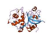
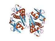
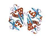
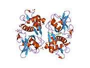
PDB gallery | ||||||||||||||||||||||||||||||||||||||||
|---|---|---|---|---|---|---|---|---|---|---|---|---|---|---|---|---|---|---|---|---|---|---|---|---|---|---|---|---|---|---|---|---|---|---|---|---|---|---|---|---|
| ||||||||||||||||||||||||||||||||||||||||