Ziehl–Neelsen stain
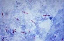
The Ziehl–Neelsen stain, also known as the acid-fast stain, was first described by two German doctors: the bacteriologist Franz Ziehl (1859–1926) and the pathologist Friedrich Neelsen (1854–1898). It is a special bacteriological stain used to identify acid-fast organisms, mainly Mycobacteria. Mycobacterium tuberculosis is the most important of this group because it is responsible for tuberculosis (TB). Other important Mycobacterium species involved in human disease are Mycobacterium leprae, Mycobacterium kansasii, Mycobacterium marinum, Mycobacterium bovis, Mycobacterium africanum and members of the Mycobacterium avium complex. Acid fast organisms like Mycobacterium contain large amounts of lipid substances within their cell walls called mycolic acids. These acids resist staining by ordinary methods such as a Gram stain.[1] It can also be used to stain a few other bacteria, such as Nocardia. The reagents used are Ziehl–Neelsen carbol fuchsin, acid alcohol, and methylene blue. Acid-fast bacilli will be bright red after staining.
A variation on this staining method is used in mycology to differentially stain acid-fast incrustations in the cuticular hyphae of certain species of fungi in the genus Russula.[2][3] It is also useful in the identification of some protozoa, namely Cryptosporidium and Isospora. The Ziehl–Neelsen stain can also hinder diagnosis in the case of paragonimiasis because the eggs in an ovum and parasite sputum sample (OnP) can be dissolved by the stain, and is often used in this clinical setting because signs and symptoms of paragonimiasis closely resemble those of TB.
Procedure
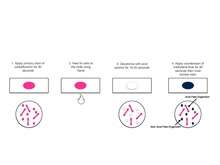
A typical AFB stain procedure involves dropping the cells in suspension onto a slide, then air drying the liquid and heat fixing the cells. The slide is flooded with Carbol Fuchsin, which is then heated to dry and rinsed off in tap water. The slide is then flooded with a 1% solution of hydrochloric acid in isopropyl alcohol (or methanol) to remove the carbol fuchsin, thus removing the stain from cells that are unprotected by a waxy lipid layer. Thereafter, the cells are stained in methylene blue and viewed on a microscope under oil immersion.
Studies have shown that an AFB stain without a culture has a poor negative predictive value. An AFB Culture should be performed along with an AFB stain; this has a much higher negative predictive value.
Mechanism explanation
Initially, Carbol Fuchsin stains every cell. When they are destained with acid-alcohol, only non-acid-fast bacteria get destained since they do not have a thick, waxy lipid layer like acid-fast bacteria. When counter stain is applied, non-acid-fast bacteria pick it up and become blue when viewed under the microscope. Acid-fast bacteria retain Carbol Fuchsin so they appear red.
Modifications
- 1% sulfuric acid alcohol for actinomycetes, nocardia.
- 0.5–1% sulfuric acid alcohol for oocysts of isospora, cyclospora.
- 0.25–0.5% sulfuric acid alcohol for bacterial endospores.
- Brucella differential stain – glacial acetic acid used, no heat applied, secondary stain is loeffler's methylene blue.
- Kinyoun modification (or cold Ziehl–Neelsen technique) is also available.
- A protocol in which a detergent is substituted for the highly toxic phenol in the fuchsin staining solution.[4]
See also
References
- "Microbiology with Diseases by Body System", Robert W. Bauman, 2009, Pearson Education, Inc.
- Morello, Josephine A., Paul A. Granato, Marion E. Wilson, and Verna Morton. Laboratory Manual and Workbook in Microbiology: Applications to Patient Car. 10th ed. Boston: McGraw-Hill Higher Education, 2006. Print.
Online protocol examples
- Ziehl–Neelsen protocol (PDF format).
References
- ↑ Morello, Josephine A., Paul A. Granato, Marion E. Wilson, and Verna Morton. Laboratory Manual and Workbook in Microbiology: Applications to Patient Care. 10th ed. Boston: McGraw-Hill Higher Education, 2006. Print.
- ↑ Romagnesi, H. (1967). Les Russules d'Europe et d'Afrique du Nord. Bordas. ISBN 0-934454-87-6.
- ↑ Largent, D; D Johnson; R Watling (1977). How to identify fungi to genus III: microscopic features. Mad River Press. ISBN 0-916422-09-7. p 25.
- ↑ Ellis, RC; LA Zabrowarny. (1993). "Safer staining method for acid fast bacilli" (PDF). Journal of Clinical Pathology. 46: 559–560. doi:10.1136/jcp.46.6.559. PMC 501296
 . PMID 7687254.
External links
-
 Media related to Ziehl-Neelsen stain at Wikimedia Commons