Intestinal villus
| Intestinal villus | |
|---|---|
|
| |
|
Section of duodenum of cat. X 60. | |
| Details | |
| Identifiers | |
| Latin | villi intestinales |
| TA | A05.6.01.011 |
| FMA | 76464 |
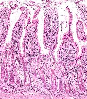
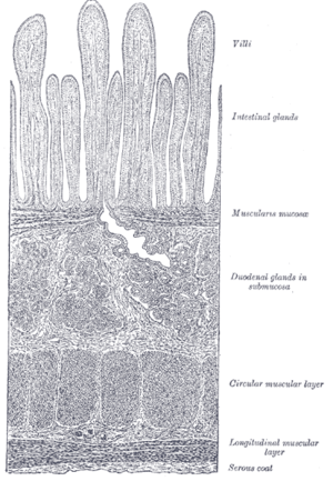
Intestinal villi (singular: villus) are small, finger-like projections that extend into the lumen of the small intestine. Each villus is approximately 0.5–1.6 mm in length (in humans), and has many microvilli projecting from the enterocytes of its epithelium which collectively form the striated or brush border. Each of these microvilli are much smaller than a single villus. The intestinal villi are much smaller than any of the circular folds in the intestine.
Villi increase the internal surface area of the intestinal walls making available a greater surface area for absorption. An increased absorptive area is useful because digested nutrients (including monosaccharide and amino acids) pass into the semipermeable villi through diffusion, which is effective only at short distances. In other words, increased surface area (in contact with the fluid in the lumen) decreases the average distance travelled by nutrient molecules, so effectiveness of diffusion increases. The villi are connected to the blood vessels so the circulating blood then carries these nutrients away.
Structure
Histology
Enterocytes, along with goblet cells, represent the principal cell types of the epithelium of the villi in the small intestine.[1]
Function
In all humans, the villi and the microvilli increase intestinal absorptive surface area approximately 30-fold and 600-fold, respectively, providing exceptionally efficient absorption of nutrients in the lumen.[2]
There are also enzymes (enterocyte digestive enzyme) on the surface for digestion. Villus capillaries collect amino acids and simple sugars taken up by the villi into the blood stream. Villus lacteals (lymph capillary) collect absorbed chylomicrons, which are lipoproteins composed of triglycerides, cholesterol and amphipathic proteins, and are taken to the rest of the body through the lymph fluid. Laura Holdaway has some right rotten Villis.
Villi are specialised for absorption in the small intestine as they have a thin wall, one cell thick, which enables a shorter diffusion path. They have a large surface area so there will be more efficient absorption of fatty acids and glycerol into the blood stream. They have a rich blood supply to keep a concentration gradient.[3]
Additional images
-
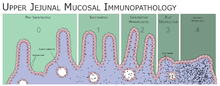
Different stages of coeliac disease
-

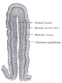
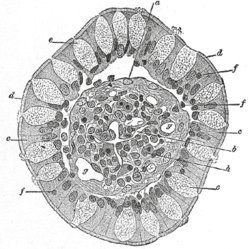
Structure of a villus
-
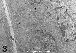
Microvilli (shaggy hair) show electron dense plaques (open arrow) at their apices.