Vaginal discharge
| Vaginal discharge | |
|---|---|
| Classification and external resources | |
| DiseasesDB | 28137 |
| MedlinePlus | 003158 |
| MeSH | D019522 |
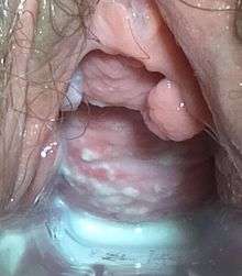
Vaginal discharge is a mixture of liquid, cells, and bacteria that lubricates the vagina.[1] This physiologic mixture is constantly produced by the cells of the vagina and cervix and exits the body through the vaginal opening. The composition, amount, and quality of discharge varies between individuals as well as through the various stages of sexual and reproductive development.[2] While most discharge represents normal functioning of the body, some changes in discharge can reflect infection or other pathological processes.[3]
Composition
The vaginal lubricative fluid contains water, pyridine, squalene, urea, acetic acid, lactic acid, complex alcohols and glycols, ketones, and aldehydes.[4] It can vary in consistency, texture, taste, color, and odor, depending on sexual arousal, the phase of the menstrual cycle, the presence of an infection, certain drugs, genetic factors, and diet.
Vaginal fluid is slightly acidic and can become more acidic with certain sexually transmitted diseases. The normal pH of vaginal fluid is between 3.8 and 4.5,[5][6] whereas male semen is typically between 7.2 and 8.0 (a neutral substance has a pH of 7.0).[7]
Production
The human vagina is serviced by nerves that respond to vasoactive intestinal polypeptide (VIP). As a consequence, VIP induces an increase in vaginal blood flow accompanied by an increase in vaginal lubrication. The findings suggest that VIP may participate in the control of the local physiological changes observed during sexual arousal: genital vasodilation and increase in vaginal lubrication.[8]
Nonpathological
Nonpathological vaginal discharge ranges in pH from 3.8 to 4.2 and is typically white or clear in color, though it can be yellowish. Nothing is visible under a microscope in a vaginal wet mount and treatment with KOH does not give off an odor, which could indicate pathology.[9][10] It is produced by cervical and vaginal glands.[9]
Menstrual cycle
Vaginal discharge amounts vary as the menstrual cycle progresses. Ovulation can increase discharge.[11] Menopause or low estrogen levels can cause a decrease in discharge.[9]
Neonatal
In neonates, vaginal discharge sometimes occurs in the first few days after birth. This is due to exposure to estrogen while in utero. Neonatal vaginal discharge may be white or clear with a mucous texture, or it may be bloody from normal transient shedding of the endometrium.[10]
In pregnancy
During pregnancy, vaginal discharge volume can increase significantly.[9]
Other factors
Vaginal discharge may increase due to stress or sexual arousal.[9]
Pathological
Pathological discharge can occur in a number of conditions, including infections and imbalances in vaginal flora or pH. Unusual vaginal discharge may also be idiopathic (the cause is not determined). Diagnosing the cause of abnormal vaginal discharge can be difficult, though a potassium hydroxide test or vaginal pH analysis may be used. When abnormal discharge occurs with burning, irritation, or itching on the vulva, it is called vaginitis.[10]
Bacterial Vaginosis
Bacterial vaginosis (BV) is an infection caused by a change in the vaginal flora, which refers to the community of organisms that live in the vagina.[12] It is the most common cause of pathological vaginal discharge in women of childbearing age and accounts for 40-50% of cases. (add cite) In BV, the vagina experiences a decrease in a bacteria called lactobacilli, and a relative increase in a multitude of anaerobic bacteria with the most predominant being Gardnerella vaginalis.[13] This imbalance results in the characteristic vaginal discharge experienced by patients with BV.[12] The discharge in BV has a characteristic strong fishy odor, which is caused by the relative increase in anaerobic bacteria.[1] The discharge is typically thin and grey, or occasionally green. It sometimes is accompanied by burning with urination. Itching is rare.[14] The exact reasons for the disruption of vaginal flora leading to BV are not fully known.[15] However, factors associated with BV include antibiotic use, unprotected sex, douching, and using an intrauterine device (IUD).[16] The role of sex in BV is unknown, and BV is not considered an STD. The diagnosis of BV is made by a health care provider based on the appearance of the discharge, discharge pH > 4.5, presence of clue cells under the microscope, and a characteristic fishy odor when the discharge is placed on a slide and combined with potassium hydroxide ("whiff test"). The gold standard for diagnosis is a gram stain showing a relative lack of lactobacilli and a polymicrobial array of gram negative rods, gram variable rods, and cocci. BV may be treated with oral or intravaginal antibiotics, or oral or intravaginal lactobacillus.[17]
Vaginal yeast infection
A vaginal yeast infection results from overgrowth of candida albicans, or yeast, in the vagina.[18] Risk factors for yeast infections include recent antibiotic use, diabetes, immunosuppression, increased estrogen levels, and use of certain contraceptive devices including intrauterine devices, diaphragms, or sponges. It is not a sexually transmitted infection. Candida vaginal infections are common; an estimated 75% of women will have at least one yeast infection in their lifetime.[19] Vaginal discharge is not always present in yeast infections, but when occurring it is typically odorless, thick, white, and clumpy. Vaginal itching is the most common symptom of candida vulvovaginitis. Women may also experience burning, soreness, irritation, pain during urination, or pain during sex.[19] The diagnosis of Candida vulvovaginitis is made by looking at a sample taken from the vagina under the microscope that shows yeast, or from a culture.[20] It is important to note that the symptoms described above may be present in other vaginal infections, so microscopic diagnosis or culture is needed to confirm the diagnosis. Treatment is with intra-vaginal or oral anti-fungal medications.
Trichomonas Vaginitis
Trichomonas vaginitis is an infection acquired through sex that is associated with vaginal discharge.[21] It can be transmitted by way of the penis to the vagina, the vagina to the penis, or from vagina to vagina.[22] The discharge in Trichomonas is typically yellowish-green in color though may also be gray or white, and may have a foul odor.[21] Other symptoms may include vaginal burning or itching, pain with urination, or pain with sexual intercourse.[22] Trichomonas is diagnosed by looking at a sample of discharge under the microscope.[21] However, in women with trichomonas the organism is typically detected in only 60-70% of cases.[21] Other testing, including a culture of the discharge or a PCR assay, are more likely to detect the organism.[21] Treatment is with a one time dose of oral antibiotics, most commonly metronidazole or tindazole.[21]
Diagnosis
Co-occurring symptoms
In bacterial vaginosis, co-occurring symptoms include a fishy odor that worsens during menstruation or after vaginal sex. In a vaginal yeast infection (candidiasis), abnormal discharge may be accompanied with itching and burning. Trichomoniasis can cause a foul odor, itching, light bleeding (spotting), and painful urination. Other bacterial infections, such as those caused by Streptococcus, Staphylococcus, or E. coli may cause itching.[10]
Color, texture, and volume
The color and texture of vaginal discharge can indicate pathology. In bacterial vaginosis, discharge can appear thin, gray, or white, increase in volume, and is adherent. In yeast infections, discharge is described as resembling cottage cheese. Trichomoniasis causes greenish-yellow, adherent, frothy discharge and can cause an increase in discharge. Bacterial infections can cause discharge containing pus.[10]
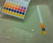
Potassium hydroxide test
The potassium hydroxide test assess whether or not a fishy odor is given off when vaginal discharge is exposed to the strong base. In bacterial vaginosis it is almost always positive, and it may be positive in trichomoniasis infections.[10]
Vaginal pH
The pH of vaginal discharge may change depending on the disease present. Normally, the pH of vaginal discharge is between 3.8 and 4.2. In aerobic vaginitis,[23] bacterial vaginosis, trichomoniasis, and bacterial infections, it is raised above 4.5, becoming more alkaline (basic). In candidiasis, it is below 4.5.[10]

Microscopic examination
A microscopic examination of vaginal discharge may indicate pathology. In candidiasis, a potassium hydroxide preparation can show hyphae and buds of the fungus. In bacterial vaginosis and trichomoniasis, a saline wet mount will show clue cells and trichomonads, respectively. Bacterial vaginosis may also cause white blood cells and clumps of bacteria to be visible in the discharge. In bacterial infections, a large number of white blood cells will be seen under the microscope.[10]
See also
References
- ↑ Beckmann, R.B. (2014). Obstetrics and Gynecology (7th ed.). Baltimore, MD: Lippincott Williams & Wilkins. p. 260. ISBN 9781451144314.
- ↑ Hacker, Neville F. (2016). Hacker & Moore's Essentials of Obstetrics and Gynecology (6th ed.). Philadelphia, PA: Elsevier. p. 276. ISBN 9781455775583.
- ↑ Lentz, Gretchen M. (2012). Comprehensive Gynecology (6th ed.). Philadelphia, PA: Elsevier. pp. 532–533. ISBN 9780323069861.
- ↑ "The-Clitoris.com: Female Body Fluids". Retrieved 2007-10-22.
- ↑ "Device and Method for Identifying and Treating Vaginal Affections". Retrieved 2007-10-18.
- ↑ Moses, Scott (2000). "Vaginal Fluid pH". Family Practice Notebook, LLC. Archived from the original on 2007-01-06. Retrieved 2007-02-04.
- ↑ "Semen analysis". Archived from the original on October 17, 2007. Retrieved 2007-10-18.
- ↑ Ottesen B, Pedersen B, Nielsen J, Dalgaard D, Wagner G, Fahrenkrug J (1987). "Vasoactive intestinal polypeptide (VIP) provokes vaginal lubrication in normal women". Peptides. 8 (5): 797–800. doi:10.1016/0196-9781(87)90061-1. PMID 3432128.
- 1 2 3 4 5 "Vaginal itching and discharge – Adult and adolescent: MedlinePlus Medical Encyclopedia". www.nlm.nih.gov. Retrieved 2015-10-30.
- 1 2 3 4 5 6 7 8 Hoffman, Barbara; Schorge, John; Schaffer, Joseph; Halvorson, Lisa; Bradshaw, Karen; Cunningham, F. (2012-04-12). Williams Gynecology, Second Edition. McGraw Hill Professional. ISBN 9780071716727.
- ↑ http://www.checkpregnancy.com/cervical-mucus-guide-ovulation-discharge/
- 1 2 al.], Alan H. DeCherney ... [et (2012). Current diagnosis & treatment : obstetrics & gynecology (11th ed. ed.). Stamford, Conn.: Appleton & Lange. ISBN 978-0071638562.
- ↑ Keane, F; Ison, C A; Noble, H; Estcourt, C (1 December 2006). "Bacterial vaginosis". Sexually Transmitted Infections. 82 (suppl_4): iv16–iv18. doi:10.1136/sti.2006.023119.
- ↑ "What are the symptoms of bacterial vaginosis?". www.nichd.nih.gov.
- ↑ "STD Facts - Bacterial Vaginosis". www.cdc.gov.
- ↑ "What causes bacterial vaginosis (BV)?". www.nichd.nih.gov.
- ↑ "The effects of antimicrobial treatment on bacterial vaginosis in non-pregnant women | Cochrane". doi:10.1002/14651858.CD006055.pub2.
- ↑ al.], editors, Richard P. Usatine ... [et (2013). The color atlas of family medicine (2nd ed. ed.). New York: McGraw-Hill. ISBN 978-0071769648.
- 1 2 Barry L. Hainer; Maria V. Gibson. "Vaginitis: Diagnosis and Treatment - American Family Physician". www.aafp.org.
- ↑ "Vulvovaginal Candidiasis - 2015 STD Treatment Guidelines". www.cdc.gov.
- 1 2 3 4 5 6 al.], editors, Richard P. Usatine ... [et (2013). The color atlas of family medicine (2nd ed. ed.). New York: McGraw-Hill. ISBN 978-0071769648.
- 1 2 "STD Facts - Trichomoniasis". www.cdc.gov. Retrieved 2016-12-04.
- ↑ Donders, Gilbert G.G.; Vereecken, Annie; Bosmans, Eugene; Dekeersmaecker, Alfons; Salembier, Geert; Spitz, Bernard (2002). "Definition of a type of abnormal vaginal flora that is distinct from bacterial vaginosis: aerobic vaginitis". BJOG 109 (1): 34–43. doi:10.1111/j.1471-0528.2002.00432.x. PMID 11845812