Uncinate fasciculus
| Uncinate fasciculus | |
|---|---|
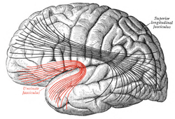 Lateral surface of left cerebral hemisphere. Some of major association tracts are depicted. Uncinate fasciculus is at lower left, in red. | |
 Human brain that operculum has been removed. A part of uncinate fasciculus is visible (shown in yellow) | |
| Details | |
| Identifiers | |
| Latin | Fasciculus uncinatus |
| NeuroNames | hier-1444 |
| NeuroLex ID | Uncinate fasciculus |
| TA | A14.1.09.560 |
| FMA | 77636 |
The uncinate fasciculus is a white matter tract in the human brain that connects parts of the limbic system such as the hippocampus and amygdala in the temporal lobe with frontal ones such as the orbitofrontal cortex. Its function is unknown though it is affected in several psychiatric conditions. It is the last white matter tract to mature in the human brain.
Anatomy
The uncinate fasciculus is a hook-shaped bundle that links the anterior portions of the temporal lobe with the inferior frontal gyrus and the lower surfaces of the frontal lobe. It does this by arising lateral to the amygdala and hippocampus in the temporal lobe curving in an upward pathway behind the external capsule inward of the insular cortex and continuing up into the posterior part of the orbital gyrus.[1]
The average length of the uncinate fasciculus is 45 mm with a range 40–49 mm. Its volume in adults is 1425.9±138.6 mm3, being slightly larger in men, at 1504.3±150.4, than women 1378.5±107.4.[2]
It has three parts: a ventral or frontal extension, an intermediary segment called the isthmus or insular segment and a temporal or dorsal segment.[3]
Function
The function of the uncinate fasciculus is not known, though it is traditionally considered to be part of the limbic system.[2]
Diffusion tensor imaging, a reconstruction model available from a diffusion MRI scan, shows a greater fractional anisotropy on the left side than on the right. The difference in this measure of anisotropy has been linked to the left hemispheric specialization for language.[4] However, the use of electrical brain stimulation upon it fails to disrupt language, suggesting it might not be involved in language, though it is possible that this disruption failed to happen because it was functionally compensated by alternative pathways.[5]
The capacity for autonoetic self-awareness that is re-experiencing previous events as part of one's past as a continuous entity across time has been linked to the right uncinate fasciculus[6] as has proficiency in auditory-verbal memory and declarative memory to the integrity of the left uncinate fasciculus.[7]
Development
The uncinate fasciculus has the longest period of development in terms of fractional anisotropy as it alone amongst the major white fibre tracks continues to develop beyond the age of 30.[8]
It seems to be developmentally vulnerable. In 12-year-old males that were preterm, abnormalities measured by fractional anisotropy in the left anterior uncinate correlated with verbal IQ, full-scale IQ, and Peabody Picture Vocabulary Test-Revised scores.[9] In 10-year-old children who have suffered socioemotional deprivation, the left uncinate fasciculus shows reduced fractional anisotropy compared to that in other children, and this might underlie their cognitive, socioemotional, and behavioral difficulties.[10]
Clinical significance
Abnormalities within the fiber bundles of the uncinate fasciculus associate with social anxiety,[11] Alzheimer's disease,[12] bipolar disorder,[13] and depression in the elderly that had previously been present in adolescence or early adulthood.[14]
Such abnormalities also link to schizophrenia.[13][15][16] In those with schizotypal personality disorder, reduced fractional anisotropy in the right uncinate fasciculus associates personality traits and clinical symptoms of ideas of reference, suspiciousness, restricted affect, reduced extraversion and social anxiety, while those on the left side associate with general intelligence, verbal and visual memory, and executive performance.[17][18] The greater left than right fractional anisotropy of the uncinate fasciculus is missing in those with schizophrenia.[19]
In 2009 it was implicated in psychopathy—individuals with a high score in the Psychopathy Checklist and an associated history of violent behavior appeared to have abnormalities in it.[20]
Phineas Gage ( a railroad worker who had an iron bar go through his left frontal lobe)[21] had damage done to his uncinate fasciculus. After the accident, his intellect was untouched, but his personality transformed. He lost all sense of morality and concern for others.
Additional images
 Diagram showing principal systems of association fibers in the cerebrum. (Uncinate fasc. visible at lower left, in red.)
References
- ↑ Kier LE, Staib LH, Davis, LM, Bronen, RA (May 1, 2004). "MR Imaging of the Temporal Stem: Anatomic Dissection Tractography of the Uncinate Fasciculus, Inferior Occipitofrontal Fasciculus, and Meyer's Loop of the Optic Radiation.". Am J Neuroradiol. 25 (5): 677–691. PMID 15140705. Retrieved 2007-12-19.
- 1 2 Hasan, KM; Iftikhar, A; Kamali, A; Kramer, LA; Ashtari, M; Cirino, PT; Papanicolaou, AC; Fletcher, JM; Ewing-Cobbs, L (2009). "Development and aging of the healthy human brain uncinate fasciculus across the lifespan using diffusion tensor tractography". Brain Research. 1276: 67–76. doi:10.1016/j.brainres.2009.04.025. PMC 2693464
 . PMID 19393229.
 - ↑ Peltier, J; Verclytte, S; Delmaire, C; Pruvo, JP; Godefroy, O; Le Gars, D (2010). "Microsurgical anatomy of the temporal stem: clinical relevance and correlations with diffusion tensor imaging fiber tracking.". Journal of Neurosurgery. 112 (5): 1033–8. doi:10.3171/2009.6.JNS08132. PMID 19612976.
- ↑ Rodrigo, S; Naggara, O; Oppenheim, C; Golestani, N; Poupon, C; Cointepas, Y; Mangin, JF; Le Bihan, D; Meder, JF.; et al. (2007). "Human subinsular asymmetry studied by diffusion tensor imaging and fiber tracking". American Journal of Neuroradiology. 28 (8): 1526–31. doi:10.3174/ajnr.A0584. PMID 17846205.
- ↑ Duffau, H; Gatignol, P; Moritz-Gasser, S; Mandonnet, E (2009). "Is the left uncinate fasciculus essential for language? A cerebral stimulation study.". Journal of neurology. 256 (3): 382–9. doi:10.1007/s00415-009-0053-9. PMID 19271103.
- ↑ Levine, B; Black, SE; Cabeza, R; Sinden, M; McIntosh, AR; Toth, JP; Tulving, E; Stuss, DT (1998). "Episodic memory and the self in a case of isolated retrograde amnesia.". Brain : a journal of neurology. 121. ( Pt 10) (10): 1951–73. doi:10.1093/brain/121.10.1951. PMID 9798749.
- ↑ Mabbott, DJ; Rovet, J; Noseworthy, MD; Smith, ML; Rockel, C (2009). "The relations between white matter and declarative memory in older children and adolescents.". Brain Research. 1294: 80–90. doi:10.1016/j.brainres.2009.07.046. PMID 19632208.
- ↑ Lebel, C; Walker, L; Leemans, A; Phillips, L; Beaulieu, C. (2008). "Microstructural maturation of the human brain from childhood to adulthood". NeuroImage. 40 (3): 1044–55. doi:10.1016/j.neuroimage.2007.12.053. PMID 18295509.
- ↑ Constable, RT; Ment, LR; Vohr, BR; Kesler, SR; Fulbright, RK; Lacadie, C; Delancy, S; Katz, KH; et al. (2008). "Prematurely born children demonstrate white matter microstructural differences at 12 years of age, relative to term control subjects: an investigation of group and gender effects". Pediatrics. 121 (2): 306–16. doi:10.1542/peds.2007-0414. PMID 18245422.
- ↑ Eluvathingal, TJ; Chugani, HT; Behen, ME; Juhász, C; Muzik, O; Maqbool, M; Chugani, DC; Makki, M. (2006). "Abnormal brain connectivity in children after early severe socioemotional deprivation: a diffusion tensor imaging study". Pediatrics. 117 (6): 2093–100. doi:10.1542/peds.2005-1727. PMID 16740852.
- ↑ Phan, KL; Orlichenko, A; Boyd, E; Angstadt, M; Coccaro, EF; Liberzon, I; Arfanakis, K (2009). "Preliminary evidence of white matter abnormality in the uncinate fasciculus in generalized social anxiety disorder.". Biological Psychiatry. 66 (7): 691–4. doi:10.1016/j.biopsych.2009.02.028. PMC 2743779. PMID 19362707.
- ↑ Yasmin, H; Nakata, Y; Aoki, S; Abe, O; Sato, N; Nemoto, K; Arima, K; Furuta, N; et al. (2008). "Diffusion abnormalities of the uncinate fasciculus in Alzheimer's disease: diffusion tensor tract-specific analysis using a new method to measure the core of the tract". Neuroradiology. 50 (4): 293–9. doi:10.1007/s00234-007-0353-7. PMID 18246334.
- 1 2 McIntosh, AM; Maniega, SM; Lymer, GK; McKirdy, J; Hall, J; Sussmann, JE; Bastin, ME; Clayden, JD; et al. (2008). "White matter tractography in bipolar disorder and schizophrenia". Biol Psychiatry. 64 (12): 1088–92. doi:10.1016/j.biopsych.2008.07.026. PMID 18814861.
- ↑ Taylor, WD; Macfall, JR; Gerig, G; Krishnan, RR. (2007). "Structural integrity of the uncinate fasciculus in geriatric depression: Relationship with age of onset". Neuropsychiatr Dis Treat. 3 (5): 669–74. PMC 2656303. PMID 19300596.
- ↑ Kubicki, M; Westin, CF; Maier, SE; Frumin, M; Nestor, PG; Salisbury, DF; Kikinis, R; Jolesz, FA; et al. (2002). "Uncinate fasciculus findings in schizophrenia: a magnetic resonance diffusion tensor imaging study". American Journal of Psychiatry. 159 (5): 813–20. doi:10.1176/appi.ajp.159.5.813. PMC 2803760. PMID 11986136.
- ↑ Kawashima, T; Nakamura, M; Bouix, S; Kubicki, M; Salisbury, DF; Westin, CF; McCarley, RW; Shenton, ME. (2009). "Uncinate fasciculus abnormalities in recent onset schizophrenia and affective psychosis: a diffusion tensor imaging study". Schizophr Res. 110 (1–3): 119–26. doi:10.1016/j.schres.2009.01.014. PMC 2749228. PMID 19328656.
- ↑ Nakamura, M; McCarley, RW; Kubicki, M; Dickey, CC; Niznikiewicz, MA; Voglmaier, MM; Seidman, LJ; Maier, SE; et al. (2005). "Fronto-temporal disconnectivity in schizotypal personality disorder: a diffusion tensor imaging study". Biol Psychiatry. 58 (6): 468–78. doi:10.1016/j.biopsych.2005.04.016. PMC 2768055. PMID 15978550.
- ↑ Gurrera, RJ; Nakamura, M; Kubicki, M; Dickey, CC; Niznikiewicz, MA; Voglmaier, MM; McCarley, RW; Shenton, ME; et al. (2007). "The uncinate fasciculus and extraversion in schizotypal personality disorder: a diffusion tensor imaging study". Schizophr Res. 90 (1–3): 360–2. doi:10.1016/j.schres.2006.10.003. PMC 1876710. PMID 17126532.
- ↑ Park, HJ; Westin, CF; Kubicki, M; Maier, SE; Niznikiewicz, M; Baer, A; Frumin, M; Kikinis, R; et al. (2004). "White matter hemisphere asymmetries in healthy subjects and in schizophrenia: a diffusion tensor MRI study". NeuroImage. 23 (1): 213–23. doi:10.1016/j.neuroimage.2004.04.036. PMC 2794419. PMID 15325368.
- ↑ Craig, Michael C; Marco Catani; Q Deeley; R Latham; E Daly; R Kanaan; M Picchioni; P K McGuire; T Fahy; Declan G M Murphy (2009-06-09). "Altered connections on the road to psychopathy". Molecular Psychiatry. 14 (10): 946–53, 907. doi:10.1038/mp.2009.40. PMID 19506560. Retrieved 2009-08-05. Lay summary – The Times.
- ↑ Horn, J Van; Irimia, A; et al. (2012). "Mapping Connectivity Damage in the Case of Phineas Gage". PLOS ONE. 7 (5): e37454. doi:10.1371/journal.pone.0037454. PMC 3353935. PMID 22616011.
External links
| Wikimedia Commons has media related to Uncinate fasciculus. |
- Atlas image: n1a5p6 at the University of Michigan Health System - "Dissection of the Left Hemisphere"