Tuberculum impar
| Unpaired swelling | |
|---|---|
 Floor of pharynx of human embryo about twenty-six days old. | |
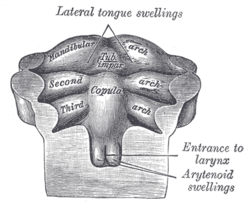 Floor of pharynx of human embryo of about the end of the fourth week. | |
| Details | |
| Gives rise to | tongue |
| Latin | tuberculum linguale mediale, tuberculum impar, tuber impar |

a Tuberculum laterale
b Tuberculum impar
c Foramen cecum
d Ductus thyreoglossus
e Sinus cervicalis
During the third week of embryogenesis there appears, immediately behind the ventral ends of the two halves of the first pharyngeal arch, a rounded swelling named the tuberculum impar or median tongue bud,[1] which was described by His as undergoing enlargement to form the buccal part of the tongue.
More recent researches, however, show that this part of the tongue is mainly, if not entirely, developed from a pair of lateral swellings which rise from the inner surface of the pharyngeal arch and meet in the middle line. The site of their meeting remains post-embryonically as the median sulcus of the tongue.
The tuberculum impar disappear in an adult tongue
References
This article incorporates text in the public domain from the 20th edition of Gray's Anatomy (1918)
External links
- hednk-024—Embryo Images at University of North Carolina