Tracheotomy
| Tracheotomy | |
|---|---|
| Intervention | |
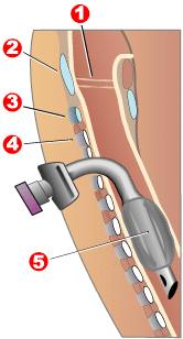 Completed tracheotomy: 1 – Vocal folds | |
| ICD-10-PCS | 0B110F4 |
| ICD-9-CM | 31.1 |
| MeSH | D014140 |
| MedlinePlus | 002955 |
Tracheotomy (US /ˌtreɪkiˈɒtəmi/ tray-kee-AW-tə-mee) is a surgical procedure which consists of making an incision on the anterior aspect of the neck and opening a direct airway through an incision in the trachea (windpipe). The resulting stoma (hole), or tracheostomy, can serve independently as an airway or as a site for a tracheostomy tube to be inserted; this tube allows a person to breathe without the use of the nose or mouth. Both surgical and percutaneous techniques are widely used in current surgical practice. It is among the oldest described procedures.
Etymology and terminology
The etymology of the word tracheotomy comes from two Greek words: the root tom- (from Greek τομή) meaning "to cut", and the word trachea (Greek τραχεία).[1] The word tracheostomy, including the root stom- (from Greek στόμα) meaning "mouth," refers to the making of a semi-permanent or permanent opening, and to the opening itself. Some sources offer different definitions of the above terms. Part of the ambiguity is due to the uncertainty of the intended permanence of the stoma at the time it is created.[2]
Indications
In the acute setting, indications for tracheotomy include such conditions as severe facial trauma, tumors of the head and neck (e.g., cancers, branchial cleft cysts), and acute angioedema and inflammation of the head and neck. In the context of failed orotracheal or nasotracheal intubation, either tracheotomy or cricothyrotomy may be performed. In the chronic setting, indications for tracheotomy include the need for long-term mechanical ventilation and tracheal toilet (e.g. comatose patients, or extensive surgery involving the head and neck). Tracheotomy may result in a significant reduction in the administration of sedatives and vasopressors, as well as the duration of stay in the intensive care unit.[3] In extreme cases, the procedure may be indicated as a treatment for severe obstructive sleep apnea (OSA) seen in patients intolerant of continuous positive airway pressure (CPAP) therapy. The reason tracheostomy works well for OSA is because it is the only surgical procedure that completely bypasses the upper airway. This procedure was commonly performed for obstructive sleep apnea until the 1980s, when other procedures such as the uvulopalatopharyngoplasty, genioglossus advancement, and maxillomandibular advancement surgeries were described as alternative surgical modalities for OSA.
Parts
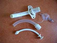
A tracheostomy tube consists of an outer cannula or main shaft, an inner cannula, and an obturator. The obturator is used when inserting the tracheostomy tube to guide the placement of the outer cannula and is removed once the outer cannula is in place. The outer cannula remains in place but, because of the buildup of secretions, there is an inner cannula that may be removed for cleaning or replaced. Tracheostomy tubes may have cuffs, inflatable balloons at the end of the tube to secure it in place. A tracheostomy tube may be fenestrated with one or several holes to let air through the larynx, allowing speech.[4]
Surgical instruments
As with most other surgical procedures, some cases are more difficult than others. Surgery on children is more difficult because of their smaller size. Difficulties such as a short neck and bigger thyroid glands make the trachea hard to open.[5] There are other difficulties with patients with irregular necks, the obese, and those with a large goitre. The many possible complications include hemorrhage, loss of airway, subcutaneous emphysema, wound infections, stomal cellulites, fracture of tracheal rings, poor placement of the tracheostomy tube, and bronchospasm".[6]
By the late 19th century, some surgeons had become proficient in performing the tracheotomy procedure. The main instruments used were:
“Two small scalpels, one short grooved director, a tenaculum, two aneurysm needles which may be used as retractors, one pair of artery forceps, haemostatic forceps, two pairs of dissecting forceps, a pair of scissors, a sharp-pointed tenotome, a pair of tracheal forceps, a tracheal dilator, tracheotomy tubes, ligatures, sponges, a flexible catheter, and feathers”.[5]Haemostatic forceps were used to control bleeding from separated vessels that were not ligatured because of the urgency of the operation. Generally, they were used to expose the trachea by clamping the isthmus thyroid gland on both sides. To open the trachea physically, a sharp-pointed tentome allowed the surgeon easily to place the ends into the opening of the trachea. The thin points permitted the doctor a better view of his incision. Tracheal dilators, such as the “Golding Bird”, were placed through the opening and then expanded by “turning the screw to which they are attached.” Tracheal forceps, as displayed on the right, were commonly used to extract foreign bodies from the larynx. The optimum tracheal tube at the time caused very little damage to the trachea and “mucus membrane”.[5]
The best position for a tracheotomy was and still is one that forces the neck into the biggest prominence. Usually, the patient was laid on his back on a table with a cushion placed under his shoulders to prop him up. The arms were restrained to ensure they would not get in the way later.[5] The tools and techniques used today in tracheotomies have come a long way. The tracheotomy tube placed into the incision through the windpipe comes in various sizes, thus allowing a more comfortable fit and the ability to remove the tube in and out of the throat without disrupting support from a breathing machine. In today’s world general anesthesia is used when performing these surgeries, which makes it much more tolerable for the patient. Special tracheostomy tube valves (such as the Passy-Muir valve[7]) have been created to assist people in their speech. The patient can inhale through the unidirectional tube. Upon expiration, pressure causes the valve to close, redirecting air around the tube, past the vocal folds, producing sound.[8]
Significant improvements to surgical instruments for tracheotomy include the direct suction tracheotomy tube invented by Josephine G. Fountain (RN); she was awarded patent no. 3039469 in 1962 for the direct suction tracheotomy tube, which improved the ways mucus could be cleared from the trachea and increased patient breathing and comfort.[9]
Percutaneous tracheotomy
While there were some earlier false starts, the first widely accepted percutaneous tracheotomy technique was described by Pat Ciaglia, a New York surgeon, in 1985. This technique involves a series of sequential dilatations using a set of seven dilators of progressively larger size.[10] The next widely used technique was developed in 1989 by Bill Griggs, an Australian intensive care specialist. This technique involves the use of a specially modified pair of forceps with a central hole enabling them to pass over a guidewire enabling the performance of the main dilation in a single step.[11] Since then a number of other techniques have been described. In 1995, Fantoni developed a translaryngeal approach of percutaneous tracheostomy which involves passing a guidewire through the larynx and over it railroading a tracheostomy tube with a cone shaped structure. It is also known as the In-and-out procedure. A variant of the original Ciaglia technique, using a single tapered dilator known as a "blue rhino", is the most commonly used of these newer techniques and has largely taken over from the early multiple dilator technique. Ambesh SP (2005) introduced a T-Trach kit (T-Dagger) which contains a T-shaped dilator with an elliptical shaft. The shaft of the dilator is marked in its length according to the sizes of tracheostomy tube to be introduced and has a number of holes. This T-shaped dilator provides better grip during its introduction and its elliptical shaft forms a calibrated tracheal stoma between two tracheal rings and minimizes tracheal ring fracture.[12] The Griggs and Ciaglia Blue Rhino techniques are the two main techniques in current use. A number of comparison studies have been undertaken between these two techniques with no clear differences emerging[13]
Complications
A 2000 Spanish study of bedside percutaneous tracheostomy reported overall complication rates of 10–15% and a procedural mortality of 0%,[14] which is comparable to those of other series reported in the literature from the Netherlands[15][16] and the United States.[17][18]
A 2003 American cadaveric study identified multiple tracheal ring fractures with the Ciaglia Blue Rhino technique as a complication occurring in 100% of their small series of cases.[19] The comparative study above also identified ring fractures in 9 of 30 live patients[13] while another small series identified ring fractures in 5 of their 20 patients.[20] The long term significance of tracheal ring fractures is unknown.
But the main complication of this procedure is obstruction of the hole due to accumulation of mucus produced by respiratory normal flora, which in turn will lead to hypoxia and further complications due to reduced level of oxygen received by lungs.[21]
The translaryngeal tracheostomy TLT Fantoni method
The translaryngeal tracheostomy is a percutaneous technique characterized by the exclusive procedure to carry out the stoma. A cone of soft plastic material, welded to a flexible cannula, is passed into trachea through the glottis, and then extracted outside of the neck through the pretracheal layers. The direction of this dilational manoeuvre is from the inside of the tracheal lumen to the outside of the neck (In/Out) and therefore completely opposite to the Out/In of other traditional percutaneous tracheostomies. The cone is then separated from the cannula, which results in it being positioned in the trachea. This method ensures considerable advantages, two of which are of particular importance: the abolition of the risk of perforation of the posterior wall and the reduction of local trauma to a level that is unlikely to be further lowered. The use of a ventilation catheter during the time of the procedure allows full control of the airway and to extend the indications of the technique to patients with severe respiratory failure. Go to website www.translaryngealtracheostomyfantoni.it for details [3].
Tracheotomy care
Caring for a tracheotomy mostly includes suctioning to prevent occlusions and replacing supplies. Because of the lack of filtering and humidifying by the nose and the ineffective cough mechanism, there is a buildup of secretions. Suctioning is only performed when clinically necessary because there are many potential risks. Risks include hypoxia and so suctioning is limited to 10 to 20 seconds at a time and the patient is hyperoxygenated just before and after suctioning. Risks also include atelectasis, or collapsing lung tissue from high suction pressure, and so pressure is limited to 80–120 mm Hg. Risks also include tissue damage. The suction catheter is inserted no more than 1 cm past the length of the tube to avoid contact with trachea tissue. Suctioning is only done during withdrawing the catheter at least 1/2 inch. Risks also include infection.[4]
Alternatives
Biphasic cuirass ventilation is a form of non-invasive mechanical ventilation that can in many cases allow patients an alternative mode of respiratory support, allowing patients to avoid an invasive tracheostomy and its many complications. While this method has not been proven to help in every case, it has been shown to be an effective alternative for many.
History
Prior to 16th century
Tracheotomy was first depicted on Egyptian artifacts in 3600 BC.[22] It was described in the Rigveda, a Sanskrit text, circa 2000 BC.[23] Homerus of Byzantium is said to have written of Alexander the Great saving a soldier from suffocation by making an incision with the tip of his sword in the man's trachea.[23] Hippocrates condemned the practice of tracheotomy as incurring an unacceptable risk of damage to the carotid artery. Warning against the possibility of death from inadvertent laceration of the carotid artery during tracheotomy, he instead advocated the practice of tracheal intubation.[6] Because surgical instruments were not sterilized at that time, infections following surgery also produced numerous complications, including dyspnea, often leading to death.[23]
Despite the concerns of Hippocrates, it is believed that an early tracheotomy was performed by Asclepiades of Bithynia, who lived in Rome around 100 BC. Galen and Aretaeus, both of whom lived in Rome in the 2nd century AD, credit Asclepiades as being the first physician to perform a non-emergency tracheotomy. Antyllus, another Roman physician of the 2nd century AD, supported tracheotomy when treating oral diseases. He refined the technique to be more similar to that used in modern times, recommending that a transverse incision be made between the third and fourth tracheal rings for the treatment of life-threatening airway obstruction.[6] Antyllus (whose original writings were lost but not before they were preserved by the Greek historian Oribasius) wrote that tracheotomy was not effective however in cases of severe laryngotracheobronchitis because the pathology was distal to the operative site.[23] In AD 131, Galen clarified the anatomy of the trachea and was the first to demonstrate that the larynx generates the voice.
By AD 700, the tracheotomy was well described in Indian and Arabian literature, although it was rarely practiced on humans.[23] In 1000, Abu al-Qasim al-Zahrawi (936–1013), an Arab who lived in Arabic Spain, published the 30-volume Kitab al-Tasrif, the first illustrated work on surgery. He never performed a tracheotomy, but he did treat a slave girl who had cut her own throat in a suicide attempt. Al-Zahrawi (known to Europeans as Albucasis) sewed up the wound and the girl recovered, thereby proving that an incision in the larynx could heal. Circa AD 1020, Avicenna (980–1037) described tracheal intubation in The Canon of Medicine in order to facilitate breathing.[24] The first correct description of the tracheotomy operation for treatment of asphyxiation was described by Ibn Zuhr (1091–1161) in the 12th century. According to Mostafa Shehata, Ibn Zuhr (also known as Avenzoar) successfully practiced the tracheotomy procedure on a goat, justifying Galen's approval of the operation.[25]
16th–18th centuries
The European Renaissance brought with it significant advances in all scientific fields, particularly surgery. Increased knowledge of anatomy was a major factor in these developments. Surgeons became increasingly open to experimental surgery on the trachea. During this period, many surgeons attempted to perform tracheotomies, for various reasons and with various methods. Many suggestions were put forward, but little actual progress was made toward making the procedure more successful. The tracheotomy remained a dangerous operation with a very low success rate, and many surgeons still considered the tracheotomy to be a useless and dangerous procedure. The high mortality rate for this operation, which had not improved, supports their position.
From the period 1500 to 1832 there are only 28 known reports of tracheotomy.[26] In 1543, Andreas Vesalius (1514–1564) wrote that tracheal intubation and subsequent artificial respiration could be life-saving. Antonio Musa Brassavola (1490–1554) of Ferrara treated a patient suffering from peritonsillar abscess by tracheotomy after the patient had been refused by barber surgeons. The patient apparently made a complete recovery, and Brassavola published his account in 1546. This operation has been identified as the first recorded successful tracheostomy, despite many ancient references to the trachea and possibly to its opening.[26] Ambroise Paré (1510–1590) described suture of tracheal lacerations in the mid-16th century. One patient survived despite a concomitant injury to the internal jugular vein. Another sustained wounds to the trachea and esophagus and died.

Towards the end of the 16th century, anatomist and surgeon Hieronymus Fabricius (1533–1619) described a useful technique for tracheotomy in his writings, although he had never actually performed the operation himself. He advised using a vertical incision and was the first to introduce the idea of a tracheostomy tube. This was a straight, short cannula that incorporated wings to prevent the tube from advancing too far into the trachea. He recommended the operation only as a last resort, to be used in cases of airway obstruction by foreign bodies or secretions. He counseled that the operation should be performed only as a last option.[23] Fabricius' description of the tracheotomy procedure is similar to that used today. Julius Casserius (1561–1616) succeeded Fabricius as professor of anatomy at the University of Padua and published his own writings regarding technique and equipment for tracheotomy. Casserius recommended using a curved silver tube with several holes in it. Marco Aurelio Severino (1580–1656), a skillful surgeon and anatomist, performed multiple successful tracheotomies during a diphtheria epidemic in Naples in 1610, using the vertical incision technique recommended by Fabricius. He also developed his own version of a trocar.[27]
In 1620 the French surgeon Nicholas Habicot (1550–1624), surgeon of the Duke of Nemours and anatomist, published a report of four successful "bronchotomies" which he had performed.[28] One of these is the first recorded case of a tracheotomy for the removal of a foreign body, in this instance a blood clot in the larynx of a stabbing victim. He also described the first tracheotomy to be performed on a pediatric patient. A 14-year-old boy swallowed a bag containing 9 gold coins in an attempt to prevent its theft by a highwayman. The object became lodged in his esophagus, obstructing his trachea. Habicot performed a tracheotomy, which allowed him to manipulate the bag so that it passed through the boy's alimentary tract, apparently with no further sequelae.[23] Habicot suggested that the operation might also be effective for patients suffering from inflammation of the larynx. He developed equipment for this surgical procedure which displayed similarities to modern designs (except for his use of a single-tube cannula).
Sanctorius (1561–1636) is believed to be the first to use a trocar in the operation, and he recommended leaving the cannula in place for a few days following the operation.[29] Early tracheostomy devices are illustrated in Habicot’s Question Chirurgicale[28] and Julius Casserius' posthumous Tabulae anatomicae in 1627.[30] Thomas Fienus (1567–1631), Professor of Medicine at the University of Louvain, was the first to use the word "tracheotomy" in 1649, but this term was not commonly used until a century later.[31] Georg Detharding (1671–1747), professor of anatomy at the University of Rostock, treated a drowning victim with tracheostomy in 1714.[32][33][34]
19th century
In the 1820s, the tracheotomy began to be recognized as a legitimate means of treating severe airway obstruction. In 1832, French physician Pierre Bretonneau employed it as a last resort to treat a case of diphtheria.[35] In 1852, Bretonneau's student Armand Trousseau reported a series of 169 tracheotomies (158 of which were for croup, and 11 for "chronic maladies of the larynx")[36] In 1858, John Snow was the first to report tracheotomy and cannulation of the trachea for the administration of chloroform anesthesia in an animal model.[37] In 1871, the German surgeon Friedrich Trendelenburg (1844–1924) published a paper describing the first successful elective human tracheotomy to be performed for the purpose of administration of general anesthesia.[38] In 1880, the Scottish surgeon William Macewen (1848–1924) reported on his use of orotracheal intubation as an alternative to tracheotomy to allow a patient with glottic edema to breathe, as well as in the setting of general anesthesia with chloroform.[39][40] At last, in 1880 Morrell Mackenzie's book discussed the symptoms indicating a tracheotomy and when the operation is absolutely necessary.[6]
20th century
In the early 20th century, physicians began to use the tracheotomy in the treatment of patients afflicted with paralytic poliomyelitis who required mechanical ventilation. However, surgeons continued to debate various aspects of the tracheotomy well into the 20th century. Many techniques were described and employed, along with many different surgical instruments and tracheal tubes. Surgeons could not seem to reach a consensus on where or how the tracheal incision should be made, arguing whether the "high tracheotomy" or the "low tracheotomy" was more beneficial. The currently used surgical tracheotomy technique was described in 1909 by Chevalier Jackson of Pittsburgh, Pennsylvania. Jackson emphasised the importance of postoperative care, which dramatically reduced the death rate. By 1965, the surgical anatomy was thoroughly and widely understood, antibiotics were widely available and useful for treating postoperative infections, and other major complications had also become more manageable.
In popular culture
- In 1973 in the 19th television season of Gunsmoke (episode 598), toward the end of the series, a backwoods community wants to hang part-time deputy and doctor-in-training, Newly O'Brian (played by Buck Taylor) for a failed emergency tracheotomy he performed. This fictional event was supposed to have occurred in the U.S. "Old West" (Kansas area) in the mid to late 1800s.
- In ninth episode of season five of the M*A*S*H* ("Mulcahy's War") in 1976, Father Mulcahy (played by William Christopher) performs an emergency tracheotomy in the field, guided by Hawkeye Pierce (played by Alan Alda) over the army two-way radio. This fictional event was supposed to have occurred during the Korean War in the early 1950s.
- In the 1975 novel Julia by Peter Straub (and in the subsequent film based on the novel), the action is precipitated when the title character attempts an emergency tracheotomy on her choking child.
- In the 2008 film Saw V, detective Peter Strahm performs an impromptu tracheotomy on himself with a ballpoint pen, preventing himself from drowning in one of the antagonist's traps.[41]
- In the pilot episode of American medical drama House, Dr Robert Chase and Dr. Allison Cameron perform a tracheostomy for a patient whose throat closes up during the MRI due to an allergic reaction to gadolinium. In the season 8 episode Twenty Vicodin (2011), the protagonist Dr. Gregory House performs an emergency tracheotomy procedure on another inmate, while in prison, using only a modified ballpoint pen he had in his hand at the time.[42]
- In the Aussie soap opera Neighbours, Karl Kennedy has to perform an emergency tracheotomy on his son Malcolm, in a 2011 episode when his windpipe is crushed.[43] In 2014, Karl talks his wife, Susan, through an emergency tracheotomy when Lou Carpenter chokes on a piece of food, while trapped under a fallen beam.[44]
- In the 2012 Australian film Daddy's Little Girl, Derek performs a tracheotomy on his brother, Tommy, to prevent him from talking.
- In the 2013 American comedy film The Heat, Sandra Bullock's character performs an unnecessary emergency tracheotomy on a choking man.[45]
- In the second episode of Malcolm in the Middle season 7, Reese stated that the Richard, the son of a really bad doctor who lets him use his equipment, gave him a tracheotomy with a crazy straw.
- In the penultimate episode of series 4 of Archer ,Sterling tries to give Pam tracheotomy with a crazy straw.
See also
References
- ↑ Romaine F. Johnson (6 March 2003). "Adult Tracheostomy". Houston, Texas: Department of Otolaryngology-Head and Neck Surgery, Baylor College of Medicine. Archived from the original on 17 May 2008.
- ↑ Jonathan P Lindman; Charles E Morgan (7 June 2010). "Tracheostomy". emedicine from WebMD. External link in
|publisher=(help) - ↑ Eberhardt, Lars Karl (2008). "Dilatational Tracheostomy on an Intensive Care Unit".
- 1 2 Taylor, C. R., Lillis, C., LeMone, P., Lynn, P. (2011) Fundamentals of nursing: The art and science of nursing care. Philadelphia: Lippincott Williams & Wilkins, page 1382-1383, 1404.
- 1 2 3 4 Wharton, Henry R. (January 1897). "Minor Surgery and Bandaging". American Journal of the Medical Sciences. 113 (1): 104–106. doi:10.1097/00000441-189701000-00008.
- 1 2 3 4 Alfio Ferlito; Alessandra Rinaldo; Ashok R. Shaha; Patrick J. Bradley (December 2003). "Percutaneous Tracheotomy". Acta Otolaryngologica. 123 (9): 1008–1012. doi:10.1080/00016480310000485. PMID 14710900.
- ↑ Passy, Victor; Baydur, A.; Prentice, W.; Darnell-Neal, R. (June 1993). "Passy-muir® tracheostomy speaking valve on ventilator-dependent patients". Laryngoscope. 103 (6): 653–658. doi:10.1288/00005537-199306000-00013. PMID 8502098.
- ↑ James H. Cullen (1 June 1963). "An Evaluation of Tracheostomy in Pulmonary Emphysema". Annals of Internal Medicine. 58 (6): 953–960. doi:10.7326/0003-4819-58-6-953. PMID 14024192.
- ↑ US patent 3039469
- ↑ Ciaglia P, Firsching R, Syniec C (June 1985). "Elective percutaneous dilatational tracheostomy. A new simple bedside procedure; preliminary report". Chest. 87 (6): 715–9. doi:10.1378/chest.87.6.715. PMID 3996056.
- ↑ Griggs WM, Worthley LI, Gilligan JE, Thomas PD, Myburg JA (June 1990). "A simple percutaneous tracheostomy technique". Surgery, Gynecology & Obstetrics. 170 (6): 543–5. PMID 2343371.
- ↑ Ambesh SP, Tripathi M, Pandey CK, Pant KC, Singh PK (July 2005). "Clinical evaluation of the 'T-Dagger': a new bedside percutaneous dilational tracheostomy device". Anaesthesia. 60 (7): 708–11. doi:10.1111/j.1365-2044.2005.04236.x. PMID 15960723.
- 1 2 Ambesh SP, Pandey CK, Srivastava S, Agarwal A, Singh DK (December 2002). "Percutaneous tracheostomy with single dilatation technique: a prospective, randomized comparison of Ciaglia blue rhino versus Griggs' guidewire dilating forceps". Anesthesia and Analgesia. 95 (6): 1739–45, table of contents. doi:10.1097/00000539-200212000-00050. PMID 12456450.
- ↑ Añón JM, Gómez V, Escuela MP, et al. (2000). "Percutaneous tracheostomy: comparison of Ciaglia and Griggs techniques". Critical Care. 4 (2): 124–8. doi:10.1186/cc667. PMC 29040
 . PMID 11056749.
 - ↑ van Heurn LW, van Geffen GJ, Brink PR (July 1996). "Clinical experience with percutaneous dilatational tracheostomy: report of 150 cases". The European Journal of Surgery. 162 (7): 531–5. PMID 8874159.
- ↑ Polderman KH, Spijkstra JJ, de Bree R, et al. (May 2003). "Percutaneous dilatational tracheostomy in the ICU: optimal organization, low complication rates, and description of a new complication". Chest. 123 (5): 1595–602. doi:10.1378/chest.123.5.1595. PMID 12740279.
- ↑ Hill BB, Zweng TN, Maley RH, Charash WE, Toursarkissian B, Kearney PA (August 1996). "Percutaneous dilational tracheostomy: report of 356 cases". The Journal of Trauma. 41 (2): 238–43; discussion 243–4. doi:10.1097/00005373-199608000-00007. PMID 8760530.
- ↑ Powell DM, Price PD, Forrest LA (February 1998). "Review of percutaneous tracheostomy". The Laryngoscope. 108 (2): 170–7. doi:10.1097/00005537-199802000-00004. PMID 9473064.
- ↑ Hotchkiss KS, McCaffrey JC (January 2003). "Laryngotracheal injury after percutaneous dilational tracheostomy in cadaver specimens". The Laryngoscope. 113 (1): 16–20. doi:10.1097/00005537-200301000-00003. PMID 12514375.
- ↑ Byhahn C, Lischke V, Halbig S, Scheifler G, Westphal K (March 2000). "Ciaglia Blue Rhino: Ein weiterentwickeltes Verfahren der perkutanen Dilatationstracheotomie" [Ciaglia blue rhino: a modified technique for percutaneous dilatation tracheostomy. Technique and early clinical results]. Der Anaesthesist (in German). 49 (3): 202–6. doi:10.1007/s001010050815. PMID 10788989.
- ↑ university of Jordan. Faculty of medicine , department of anesthesia.
- ↑ Steven E. Sittig; James E. Pringnitz (February 2001). "Tracheostomy: evolution of an airway" (PDF). AARC Times: 48–51.
- 1 2 3 4 5 6 7 O. Rajesh; R. Meher (2006). "Historical Review Of Tracheostomy". The Internet Journal of Otorhinolaryngology. 4 (2). ISSN 1528-8420.
- ↑ Patricia Skinner (2008). "Unani-tibbi". In Laurie J. Fundukian. The Gale Encyclopedia of Alternative Medicine (3rd ed.). Farmington Hills, Michigan: Gale Cengage. ISBN 978-1-4144-4872-5. External link in
|publisher=(help) - ↑ Mostafa Shehata (April 2003). "The Ear, Nose and Throat in Islamic Medicine" (PDF). Journal of the International Society for the History of Islamic Medicine. 2 (3): 2–5. ISSN 1303-667X.
- 1 2 Goodall, E.W. (1934). "The story of tracheostomy". British Journal of Children's Diseases. 31: 167–76, 253–72.
- ↑ Armytage WHG (1960). "Nova et Vetera". British Medical Journal. 1 (5179): 1129–1130. doi:10.1136/bmj.1.5179.1129. PMC 1966956.
- 1 2 Nicholas Habicot (1620). Question chirurgicale par laquelle il est démonstré que le Chirurgien doit assurément practiquer l'operation de la Bronchotomie, vulgairement dicte Laryngotomie, ou perforation de la fluste ou du polmon (in French). Paris: Corrozet. p. 108.
- ↑ Sanctorii Sanctorii (1646). Sanctorii Sanctorii Commentaria in primum fen, primi libri canonis Avicennæ (in Latin). Venetiis: Apud Marcum Antonium Brogiollum. p. 1120. OL 15197097M.
- ↑ Julius Casserius (Giulio Casserio) and Daniel Bucretius (1632). Tabulae anatomicae LXXIIX ... Daniel Bucretius ... XX. que deerant supplevit & omnium explicationes addidit (in Latin). Francofurti: Impensis & coelo Matthaei Meriani.
- ↑ Cawthorne T, Hewlett AB, Ranger D (1959). "Discussion: Tracheostomy To-Day". Proceedings of the Royal Society of Medicine. 52 (6): 403–405. PMC 1871130. PMID 13667911.
- ↑ Georges Detharding (1745). "De methodo subveniendi submersis per laryngotomiam (1714)". In Von Ernst Ludwig Rathlef; Gabriel Wilhelm Goetten; Johann Christoph Strodtmann. Geschichte jetzlebender Gelehrten, als eine Fortsetzung des Jetzlebenden. Zelle: Berlegts Joachim Undreas Deek. p. 20.
- ↑ Price JL (1962). "THE EVOLUTION OF BREATHING MACHINES". Medical History. 6 (1): 67–72. doi:10.1017/s0025727300026867. PMC 1034674. PMID 14488739.
- ↑ Wischhusen HG, Schumacher GH (1977). "Curriculum vitae des Rostocker Professors für Anatomie, Botanik und der höheren Mathematik" [Curriculum vitae of the professor of anatomy, botany and higher mathematics Georg Detharding (1671–1747) at the University of Rostock]. Anatomischer Anzeiger (in German). 142 (1–2): 133–40. PMID 339777.
- ↑ Armand Trousseau (1833). "Mémoire sur un cas de tracheotomie pratiquée dans la période extrème de croup". Journal des connaissances médico-chirurgicales. 1 (5): 41.
- ↑ Armand Trousseau (1852). "Nouvelles recherches sur la trachéotomie pratiquée dans la période extrême du croup". In Jean Lequime and J. de Biefve. Annales de médecine belge et étrangère. Brussels: Imprimerie et Librairie Société Encyclographiques des Sciences Médicales. pp. 279–288.
- ↑ Snow, J (1858). "Fatal cases of inhalation of chloroform, Treatment of suspended animation from chloroform". In Richardson, BW. On chloroform and other anaesthetics: their action and administration. London: John Churchill. pp. 120–200, 251–62.
- ↑ Trendelenburg, F (1871). "Beiträge zu den Operationen an den Luftwegen" [Contributions to airways surgery]. Archiv für Klinische Chirurgie (in German). 12: 112–33.
- ↑ Macewen, W (1880). "General Observations on the Introduction of Tracheal Tubes by the Mouth, Instead of Performing Tracheotomy or Laryngotomy". British Medical Journal. 2 (1021): 122–4. doi:10.1136/bmj.2.1021.122. PMC 2241154. PMID 20749630.
- ↑ Macewen, W (1880). "Clinical Observations on the Introduction of Tracheal Tubes by the Mouth, Instead of Performing Tracheotomy or Laryngotomy". British Medical Journal. 2 (1022): 163–5. doi:10.1136/bmj.2.1022.163. PMC 2241109. PMID 20749636.
- ↑ "Saw V". IFC. Retrieved 8 June 2014.
- ↑ Barnett, Barbara (September 15, 2011). "TV Preview: House, M.D. – Season Eight Premiere "Twenty Vicodin"". Blogcritics. Retrieved October 1, 2011.
- ↑ "Benjie's back – for a bit". Channel 5. 2 June 2011. Retrieved 2 September 2014.
- ↑ "Deadly storm". TV Soap (16): 57. 28 August – 11 September 2014.
- ↑ "The Heat (2013)". Dove. Retrieved 8 June 2014.
External links
| Wikimedia Commons has media related to Tracheotomy. |
| Look up tracheotomy, pharyngotomy, laryngotomy, or tracheostomy in Wiktionary, the free dictionary. |
- Plotnikow GA, Roux N, Feld V, et al. (October 2013). "Evaluation of tracheal cuff pressure variation in spontaneously breathing patients". International Journal of Critical Illness and Injury Science. 3 (4): 262–8. doi:10.4103/2229-5151.124148. PMC 3891193. PMID 24459624.
- Tracheotomy Info (A Community For Tracheotomy-wearers and the people who love them) at tracheotomy.info
- Tracheostomy Products and Support (Online resource for tracheostomy products, supplies and support) at trachs.com
- Aaron's tracheostomy page (Caring for a tracheostomy) at tracheostomy.com
- (Pictures with video clipping) at drtbalu.com
- "Tracheotomy" at Dorland's Medical Dictionary
- Smiths Medical Tracheostomy Training Videos
- A Video of Rescue Breathing for Laryngectomees and Neck Breathers
- "Book of Simplification Concerning Therapeutics and Diet", is a manuscript from 1497 that discusses tracheotomies
- Site and blog with information about tracheostomies
- Global Tracheostomy Collaborative. International collaborative with resources for hospitals, caregivers, and patients about tracheostomies, including international research
- Dilatational Tracheostomy On An Intensive Care Unit