Tetanospasmin


Tetanus toxin is an extremely potent neurotoxin produced by the vegetative cell of Clostridium tetani[1] in anaerobic conditions, causing tetanus. It has no known function for clostridia in the soil environment where they are normally encountered. It is also called spasmogenic toxin, or TeNT. The LD50 of this toxin has been measured to be approximately 1 ng/kg, making it second only to Botulinum toxin as the deadliest toxin in the world.[2] However, these tests are conducted solely on mice which may react to the toxin differently from humans and other animals.
C. tetani also produces the exotoxin tetanolysin, a hemolysin, that causes destruction of tissues.[3]
Distribution
Tetanus toxin spreads through tissue spaces into the lymphatic and vascular systems. It enters the nervous system at the neuromuscular junctions and migrates through nerve trunks and into the central nervous system (CNS) by retrograde axonal transport by using dyneins.[4][5]
Structure
The tetanus toxin protein has a molecular weight of 150kDa. It is translated from the TetX gene as one protein which is subsequently cleaved into two parts: a 100kDa heavy or B-chain and a 50kDa light or A-chain. The chains are connected by a disulfide bond.
- The B-chain binds to dissialogangliosides (GD2 and GD1b) on the neuronal membrane and contains a translocation domain which aids the movement of the protein across that membrane and into the neuron.
- The A-chain, a zinc endopeptidase, attacks the vesicle-associated membrane protein (VAMP).
The TetX gene encoding this protein is located on the PE88 plasmid.[6][7]
The sequence of this protein has been deposited in Uniprot with accession P04958
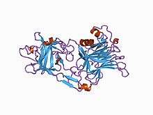
Several structures of the binding domain and the peptidase domain have been solved by X-ray crystallography and deposited in the PDB. A summary of these structures is available using the UniPDB application at PDBe, for example 1z7h or 3hmy.
Mechanism
The mechanism of TeNT action can be broken down and discussed in 6 different steps.
- Transport
- Specific binding in the periphery neurons
- Retrograde axonal transport to the central nervous system (CNS) inhibitory interneurons
- Transcytosis from the axon into the inhibitory interneurons
- Action
- Temperature and pH mediated translocation of the light chain into the cytosol
- Reduction of the disulphide bond between the light and heavy chain
- Cleavage of synaptobrevin
The first three steps outline the travel of tetanus from the peripheral nervous system to where it is taken up to the CNS and has its final effect. The last three steps document the changes necessary for the final mechanism of the neurotoxin.
Transport to the CNS inhibitory interneurons begins with The B-chain mediating the neurospecific binding of TeNT to the nerve terminal membrane. It binds to GT1b polysialogangliosides, similarly to the botulinum neurotoxin. It also binds another poorly characterized GPI anchored protein receptor more specific to TeNT.[8] [9] Both the ganglioside and the GPI anchored protein are located in lipid microdomains and both are requisite for specific TeNT binding.[9] Once it is bound the neurotoxin is then endocytosed into the nerve and begins to travel through the axon to the spinal neurons. The next step, transcytosis from the axon into the CNS inhibitory interneuron, is one of the least understood parts of TeNT action. At least two pathways are involved, one that relies on the recycling of synaptic vesicle 2 (SV2) system and one that does not.[10]
Once the vesicle is in the inhibitory interneuron its translocation is mediated by pH and temperature, specifically a low or acidic pH in the vesicle and standard physiological temperatures.[11] [12] Once the toxin has been translocated into the cytosol the disulfide bond is reduced, mainly by the NADPH-thioredoxin reductase-thioredoxin redox system and the light chain is free to cleave the Gln76-Phe77 bond of synaptobrevin.[13] Cleavage of synaptobrevin affects the stability of the SNARE core by restricting it from entering the low energy conformation which is the target for NSF binding.[14] Synaptobrevin is an integral V-SNARE necessary for vesicle fusion to membranes. The cleavage of synaptobrevin is the final target of TeNT and even in low doses the neurotoxin will inhibit neurotransmitter exocytosis in the inhibitory interneurons. The blockage of these neurotransmitters is what causes the physiological effects that accompany TeNT, specifically the blockage of the neurotransmitters GABA and glycine.
Tetanus toxin causes violent spastic paralysis by blocking the release of γ-aminobutyric acid (GABA). GABA is a neurotransmitter that inhibits motor neurons.[15]
The action of the A-chain stops the affected neurons from releasing the inhibitory neurotransmitters GABA and glycine, but also excitatory transmitters,[16] by degrading the protein synaptobrevin 2.[17] The consequence of this is dangerous overactivity in the muscles from the smallest stimulus—the failure of inhibition of motor reflexes by sensory stimulation. This causes generalized contractions of the agonist and antagonist musculature, termed a tetanic spasm.
Clinical significance
Tetanic spasms can occur in a distinctive form called opisthotonos and be sufficiently severe to fracture long bones. The shorter nerves are the first to be inhibited, which leads to the characteristic early symptoms in the face and jaw, risus sardonicus and lockjaw.
The toxin bind to the neurons is irreversible[4] and nerve function can only be returned by the growth of new terminals and synapses.
Immunity and vaccination
Due to its extreme potency, even a lethal dose of tetanospasmin may be insufficient to provoke an immune response. Naturally-acquired tetanus infections thus do not usually provide immunity to subsequent infections. Immunization (which is impermanent and must be repeated periodically) instead utilizes the less deadly toxoid derived from the toxin, as in the tetanus vaccine and some combination vaccines (such as DTP).
References
- ↑ "tetanospasmin" at Dorland's Medical Dictionary
- ↑ "Toxins of Biological Origin". Retrieved 21 November 2012.
- ↑ Willey, Joanne (2009). Prescott's Principles of Microbiology. New York City, NY: McGraw-Hill. p. 481. ISBN 978-0-07-337523-6.
- 1 2 Farrar JJ; Yen LM; Cook T; Fairweather N; Binh N; Parry J; Parry CM (September 2000). "Tetanus". Journal of Neurology, Neurosurgery, and Psychiatry. 69 (3): 292–301. doi:10.1136/jnnp.69.3.292. PMC 1737078
 . PMID 10945801.
 - ↑ AU Lalli G, Gschmeissner S, Schiavo G (November 15, 2003). "Myosin Va and microtubule-based motors are required for fast axonal retrograde transport of tetanus toxin in motor neurons". Journal of Cell Science. 116 (22): 4639–50. doi:10.1242/jcs.00727. PMID 14576357.
- ↑ Tetanus toxin: primary structure, expression in E. coli, and homology with botulinum toxins. Eisel U. et al EMBO J. 5:2495-2502(1986) PubMed: 3536478
- ↑ Popp D, Narita A, Lee LJ, Ghoshdastider U, Xue B, Srinivasan R, Balasubramanian MK, Tanaka T, Robinson RC (2012). "Novel actin-like filament structure from Clostridium tetani". The Journal of Biological Chemistry. 287 (25): 21121–9. doi:10.1074/jbc.M112.341016. PMC 3375535. PMID 22514279.
- ↑ Munro, P; Kojima, H; Dupont, JL; Bossu, JL; Poulain, B; Boquet, P (30 November 2001). "High sensitivity of mouse neuronal cells to tetanus toxin requires a GPI-anchored protein.". Biochemical and Biophysical Research Communications. 289 (2): 623–9. doi:10.1006/bbrc.2001.6031. PMID 11716521.
- 1 2 Winter, A; Ulrich, WP; Wetterich, F; Weller, U; Galla, HJ (17 June 1996). "Gangliosides in phospholipid bilayer membranes: interaction with tetanus toxin.". Chemistry and physics of lipids. 81 (1): 21–34. doi:10.1016/0009-3084(96)02529-7. PMID 9450318.
- ↑ Yeh, FL; Dong, M; Yao, J; Tepp, WH; Lin, G; Johnson, EA; Chapman, ER (24 November 2010). "SV2 mediates entry of tetanus neurotoxin into central neurons." (PDF). PLoS Pathogens. 6 (11): e1001207. doi:10.1371/journal.ppat.1001207. PMC 2991259. PMID 21124874.
- ↑ Pirazzini, M; Rossetto, O; Bertasio, C; Bordin, F; Shone, CC; Binz, T; Montecucco, C (4 January 2013). "Time course and temperature dependence of the membrane translocation of tetanus and botulinum neurotoxins C and D in neurons.". Biochemical and Biophysical Research Communications. 430 (1): 38–42. doi:10.1016/j.bbrc.2012.11.048. PMID 23200837.
- ↑ Burns, JR; Baldwin, MR (8 August 2014). "Tetanus neurotoxin utilizes two sequential membrane interactions for channel formation.". The Journal of Biological Chemistry. 289 (32): 22450–8. doi:10.1074/jbc.m114.559302. PMID 24973217.
- ↑ Pirazzini, M; Bordin, F; Rossetto, O; Shone, CC; Binz, T; Montecucco, C (16 January 2013). "The thioredoxin reductase-thioredoxin system is involved in the entry of tetanus and botulinum neurotoxins in the cytosol of nerve terminals.". FEBS Letters. 587 (2): 150–5. doi:10.1016/j.febslet.2012.11.007. PMID 23178719.
- ↑ Pellegrini, LL; O'Connor, V; Lottspeich, F; Betz, H (2 October 1995). "Clostridial neurotoxins compromise the stability of a low energy SNARE complex mediating NSF activation of synaptic vesicle fusion.". The EMBO Journal. 14 (19): 4705–13. PMID 7588600.
- ↑ Kumar, Vinay; Abbas, Abul K.; Fausto, Nelson; Aster, Jon (2009-05-28). Robbins and Cotran Pathologic Basis of Disease, Professional Edition: Expert Consult - Online (Robbins Pathology) (Kindle Locations 19359-19360). Elsevier Health. Kindle Edition.
- ↑ Kanda K, Takano K (February 1983). "Effect of tetanus toxin on the excitatory and the inhibitory post-synaptic potentials in the cat motoneurone.". J Physiol. 335: 319–333. doi:10.1113/jphysiol.1983.sp014536. PMC 1197355. PMID 6308220.
- ↑ Schiavo G, Benfenati F, Poulain B, Rossetto O, Polverino de Laureto P, DasGupta BR, Montecucco C (October 29, 1992). "Tetanus and botulinum-B neurotoxins block neurotransmitter release by proteolytic cleavage of synaptobrevin". Nature. 359 (6398): 832–5. doi:10.1038/359832a0. PMID 1331807.
External links
- tetanospasmin at the US National Library of Medicine Medical Subject Headings (MeSH)
- Tetanus and botulinum neurotoxins: mechanism of action and therapeutic uses.
- Neurotoxin in Toxicon
- The journey of tetanus and botulinum neurotoxins in neurons
- How do tetanus and botulinum toxins bind to neuronal membranes?
| Bacterial toxins |
| ||||||||||||||||||||||||||||||||||||||||||
|---|---|---|---|---|---|---|---|---|---|---|---|---|---|---|---|---|---|---|---|---|---|---|---|---|---|---|---|---|---|---|---|---|---|---|---|---|---|---|---|---|---|---|---|
| Mycotoxins |
| ||||||||||||||||||||||||||||||||||||||||||
| Plant toxins |
| ||||||||||||||||||||||||||||||||||||||||||
| Invertebrate toxins | |||||||||||||||||||||||||||||||||||||||||||
| Vertebrate toxins |
| ||||||||||||||||||||||||||||||||||||||||||
| |||||||||||||||||||||||||||||||||||||||||||
| |||||||||||||||||||||||||||||||||||||||||||