Superior gluteal nerve
| Superior gluteal nerve | |
|---|---|
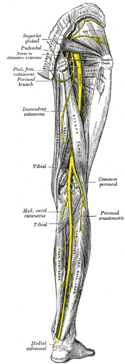 Nerves of the right lower extremity. Posterior view. | |
 Plan of sacral and pudendal plexuses. (Superior gluteal labeled at upper left.) | |
| Details | |
| From | sacral plexus (L4-S1) |
| Innervates | gluteus medius, gluteus minimus, tensor fasciæ latæ |
| Identifiers | |
| Latin | nervus gluteus superior |
| TA | A14.2.07.031 |
| FMA | 16510 |
The superior gluteal nerve is a nerve that originates in the pelvis and supplies the gluteus medius, the gluteus minimus and the tensor fasciae latae muscle.[1]
Structure
The superior gluteal nerve originates in the sacral plexus. It arises from the dorsal divisions of the L4, L5 and S1.[2] It leaves the pelvis through the greater sciatic foramen above the piriformis, accompanied by the superior gluteal artery and the superior gluteal vein.[3] It then accompanies the upper branch of the deep division of the superior gluteal artery and ends in the gluteus minimus and tensor fasciae latae muscle.
Pathology
In normal gait, the small gluteal muscles on the stance side can stabilize the pelvis in the coronal plane. Weakness or paralysis of these muscles caused by a damaged superior gluteal nerve can result in a weak abduction in the affected hip joint. This gait disturbance is known as Trendelenburg gait. In a positive Trendelenburg's sign the pelvis sags toward the normal unsupported side (the swing leg). The opposite, when the pelvis is elevated on the swing side, is known as Duchenne limp. Bilateral loss of the small gluteal muscles results in a waddling gait. [3]
See also
Notes
References
This article incorporates text in the public domain from the 20th edition of Gray's Anatomy (1918)
- Platzer, Werner (2004). Color Atlas of Human Anatomy, Vol. 1: Locomotor System (5th ed.). Thieme. ISBN 3-13-533305-1.
- Thieme Atlas of Anatomy: General Anatomy and Musculoskeletal System. Thieme. 2006. ISBN 1-58890-419-9.
External links
- Superior_gluteal_nerve at the Duke University Health System's Orthopedics program