Splanchnic nerves
| Splanchnic nerves | |
|---|---|
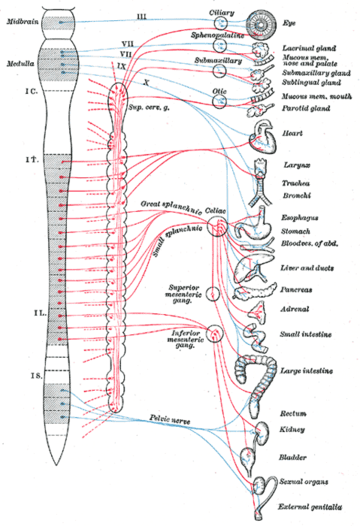 Nerves of the autonomic nervous system, with splanchnic nerves seen in center. | |
The splanchnic nerves are paired visceral nerves (nerves that contribute to the innervation of the internal organs), carrying fibers of the autonomic nervous system (visceral efferent fibers) as well as sensory fibers from the organs (visceral afferent fibers). All carry sympathetic fibers except for the pelvic splanchnic nerves, which carry parasympathetic fibers.
Types
The term splanchnic nerves can refer to:
- Cardiopulmonary nerves[1]
- Thoracic splanchnic nerves (greater, lesser, and least)
- Lumbar splanchnic nerves
- Sacral splanchnic nerves
- Pelvic splanchnic nerves
| Nerve | Pre-/postsynaptic[2] | autonomic system[2] | Origin[2] | Targets[2] | |
|---|---|---|---|---|---|
| Cardiopulmonary nerves | Postsynaptic | sympathetic | cervical and upper thoracic ganglia | Thoracic cavity | |
| Thoracic splanchnic nerves | generally | Presynaptic | lower thoracic ganglia | Prevertebral ganglia | |
| Greater splanchnic nerve | T5-T9 or T10 | Celiac ganglia | |||
| Lesser splanchnic nerve | T10-T11 | Superior mesenteric ganglia and Aorticorenal ganglia | |||
| Least splanchnic nerve | T12 | Renal plexus | |||
| Lumbar splanchnic nerves | L1-2 | Inferior mesenteric ganglia, ganglia of intermesenteric and hypogastric plexuses | |||
| Sacral splanchnic nerves | sacral part of sympathetic trunk | inferior hypogastric plexus and ganglia to the pelvic viscera | |||
| Pelvic splanchnic nerves | parasympathetic | S2-S4 | intrinsic ganglia of descending and sigmoid colon, rectum, and inferior hypogastric plexus and ganglia to the pelvic viscera | ||
References
This article is issued from Wikipedia - version of the 6/30/2016. The text is available under the Creative Commons Attribution/Share Alike but additional terms may apply for the media files.