Serratia
| Serratia | |
|---|---|
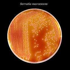 | |
| Serratia marcescens, a typical species, on XLD agar.[1] | |
| Scientific classification | |
| Domain: | Bacteria |
| Phylum: | Proteobacteria |
| Class: | Gammaproteobacteria |
| Order: | Enterobacteriales |
| Family: | Enterobacteriaceae |
| Genus: | Serratia Bizio 1823 |
| Species | |
|
S. aquatilis[2] | |
Serratia is a genus of Gram-negative, facultatively anaerobic, endosporeforming,[3] rod-shaped bacteria of the Enterobacteriaceae family. The most common and pathogenic of the species in the genus, S. marcescens, is normally the only pathogen and usually causes nosocomial infections. However, rare strains of S. plymuthica, S. liquefaciens, S. rubidaea, and S. odoriferae have caused diseases through infection.[4] S. marcescens is typically found in showers, toilet bowls, and around wetted tiles. Members of this genus produce characteristic red pigment, prodigiosin, and can be distinguished from other members of the Enterobacteriaceae family by their unique production of three enzymes: DNase, lipase, and gelatinase.[5] A pan-genome analysis of the genus Serratia revealed that it does not depict exceptional genetic conservation or diversity as compared to other bacterial genuses.[6]
Infection of humans
The bacterium is an opportunistic human pathogen, capitalizing on its ability to form tight-knit surface communities called biofilms wherever it can.[3] S. marcescens is thought to be transmitted through hand-to-hand transmission by hospital personnel.[3] In the hospital, Serratia species tend to colonize the respiratory and urinary tracts, rather than the gastrointestinal tract, in adults. Serratia infection is responsible for about 2% of nosocomial infections of the bloodstream, lower respiratory tract, urinary tract, surgical wounds, and skin and soft tissues in adult patients. Outbreaks of S. marcescens meningitis, wound infections, and arthritis have occurred in pediatric wards.[7]
Cases of Serratia arthritis have been reported in outpatients receiving intra-articular injections.
Isolation and identification of S. marcescens
Several methods can be used to study the epidemiology of Serratia marcescens. Serological typing and different types of Polymerase Chain Reactions (PCR) can be used to identify the bacteria. Biotyping, bacteriocin typing, phage typing, plasmid analysis, and ribotyping can also be used[8] Serratia marcescens appears red on Trypticase Soy Agar slants when grown at around 25 degrees Celscius[9] S. marcescensand S. liquefaciens can be easily confused in the lab when using the API system. They can both oxidise arabinose, but only S. liquefaciens can ferment arabinose in peptone water[8]
History
S. marcescens was first documented as a red coloured putrefaction of polenta[10] by Bartolomeo Bizio in Padua. The bacterium was later named in honour of Italian physicist Serafino Serrati and marcescens because of the pigment's rapid discolouration and decay.[10]:538
References
- ↑ Images courtesy of CDC Accessed 7 July 2011.
- 1 2 3 4 5 LPSN bacterio.net
- 1 2 3 Basilio J Anía (21 October 2015). "Serratia". Medscape.
- ↑ Basilio J. Anía, M.D. "Serratia". eMedicine. Retrieved 2007-03-14.
- ↑ "Serratia". University of Texas at Houston Medical School. Archived from the original on 2007-01-28. Retrieved 2007-03-14.
- ↑ . Basharat Z, Yasmin A. 2016. arXiv Preprint. arXiv: 1610.04160
- ↑ Health Canada. MSDS - Infectious Substances. Serratia. (http://www.phac-aspc.gc.ca/lab-bio/res/psds-ftss/msds138e-eng.php) Accessed 7 July 2011.
- 1 2 . HEJAZI A, FALKINER F. J. Med. Microbiol. 46(11):903-912 doi:10.1099/00222615-46-11-903
- ↑ .Leboffe, Michael J., and Burton E. Pierce. "Section 3: Bacterial Growth." A Photographic Atlas for the Microbiology Laboratory. 4th ed. Englewood, CO: Morton Pub., 2011. 26. Print.
- 1 2 "Bartolomeo Bizio's Letter to the most Eminent Priest, Angelo Bellani, Concerning the Phenomenon of the Red Colored Polenta". Journal of Bacteriology. November 1924. pp. 527–543. Retrieved 18 April 2016.