Scalp reconstruction
Scalp reconstruction is a surgical procedure for people with scalp defects. Scalp defects may be partial or full thickness and can be congenital or acquired. Because not all layers of the scalp are elastic and the scalp has a convex shape, the use of primary closure is limited. Sometimes the easiest way of closing the wound may not be the ideal or best way. The choice for a reconstruction depends on multiple factors, such as the defect itself, the patient characteristics and surgeon preference.
History

_2010-01-18.jpg)
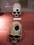
Skull and brain 'surgery' are known from the prehistoric era. There is even evidence that scalp reconstructions dates back to the Egyptians in 3000 B.C. and it was also known by the Roman Empire. The word plastic surgery probably comes from the Greek word "Plastikos" what means "shape" or "reconstruct". Damaged skulls have been tried to reconstruct, even though knowledge about neurology, the brain and anatomy were minimal. In musea skulls can be found which show manipulation that can be interpreted as a primitive form of surgery. In Medieval times, people were convinced that trepanation was a remedy for various diseases.
Indications
Main reasons for scalp reconstruction are divided into two groups: congenital or acquired. Congenital defects may include aplasia cutis congenita, congenital nevus, congenital vascular malformations and congenital tumors. Acquired defects can be caused by burns, blunt, penetrating, or avulsion injuries, tumor invasion, infection, oncologic resection, radiation, or wound-healing difficulties.[1] Alopecia can be an aesthetic motivation for hair bearing scalp reconstruction.[2] As the incidence of basal-cell carcinoma and squamous-cell carcinoma is rising and about 80% are located in the head and neck area, the number of scalp reconstructions will likely increase in the future. Depending on the size and nature of the defect, an appropriate reconstructive method has to be used. By using Mohs surgery the defect can be kept minimal, but nevertheless infiltrative basal cell carcinoma may have the need to remove a large part of the scalp.
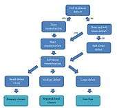
On the flowchart a simplified algorithm for scalp reconstruction is depicted. The options range from simple solutions for small skin defects to complicated reconstructions requiring multi-tissue reconstructions.
In case of an active and severe infection, it has to be controlled first by surgical debridement and antibiotic treatment before reconstruction is performed, as infection can cause bacteraemia and has a negative effect on wound healing.
Surgical anatomy
The five layers of the scalp, from superficial to deep, can be easily memorized by using the mnemonic SCALP. The Skin of the scalp is the thickest of the human body, measuring up to 8 mm in thickness and contains approximately 100.000 hairs. Hair lines make scalp reconstruction difficult because the hair lines have to be respected to get a satisfying aesthetic result.[3] The subCutis is a layer of fat, enclosed in compartments formed by rigid fibrous septa. Their inelasticy prevents bleeding vessels to collapse and retract under the skin to achieve haemostasis. All large blood vessels and nerves of the scalp are located in this layer.[4] The next layer is the galea Aponeurotica, which separates the underlying bone and the overlying layers. The large blood vessels and nerves of the scalp don’t pierce this layer.[3] Loose connective tissue between the periostium and the aponeurosis makes these two rigid structures easily slide over each other and contributes to skin movement. Thus, if vascular and nervous anatomy is respected, the skin, sucutaneous tissue and galea aponeurotica can be lifted off the skull with minimal bleeding, nerve damage, or chance of necrosis. This method was first described by Orticochea in 1967, but has been updated to minimize scarring.[5] The fifth layer is the Periosteum of the skull, also referred to as pericranium. It can be separated from the skull, except near the sutures. The skull consists of an inner and outer table, with spongy bone in between known as diploë.[4]
Vascular supply
On both sides of the scalp there are five large arteries which perfuse the scalp. Local flaps used for scalp reconstruction must contain at least one of these major arteries, to maintain a reliable blood supply. The scalp can be divided into four different vascular territories:
- Anterior: supratrochlear artery and supraorbital artery
- Lateral: superficial temporal artery
- Posterior: occipital artery
- Posterolateral: posterior auricular artery[3][6]
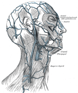

The veins anastomose frequently with each other and enter the diploic veins of the skull bones and the dural sinuses. This is an extra difficulty as the vein pattern differs. The scalp veins accompany the arteries and have similar names:
- Anterior: Supratrochlear vein and supraorbital vein
- Lateral: Superficial temporal vein
- Posterior: Occipital vein
- Posterolateral: Posterior auricular vein
Lymphatic system
The frontal part of the scalp is drained to the parotid, submandibular, and deep cervical lymph nodes. The posterior part is drained to the posterior auricular and occipital lymph nodes. Malignancies of the scalp can metastasize to these lymph nodes. Brain tumors, however, tend to metastasize haematogenously.
Innervation
The scalp is innervated by motor nerves and sensory nerves. The trigeminal nerve (CNV) is one of the important cranial sensory nerves which innervates the scalp. From anterior to posterior front to back the nerves are:
- Supratrochlear nerve and Supraorbital nerve
- Zygomaticotemporal nerve
- Auriculotemporal nerve
- Lesser occipital nerve
- Greater occipital nerve
Non-skin reconstruction
Dura mater reconstruction
Dural lesions should be closed to avoid CSF leakage. Also, a defect acts like a port of entry for micro-organisms that can cause meningitis. If fibrin glue or primary closure is not possible, patches have to be used. These are made from cadaveric dura mater, xenografts (tachosil, duragen, durepair), or synthetic grafts materials (PTFE, neuropatch). However, (vascularised) autografts (fascia lata, muscle or omentum majus) are preferred in irradiated or severely infected defects.[7]
Bone defects
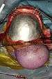
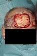
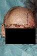
Skull defects should be closed in order to protect the brain. The occipital and temporal regions bear the most pressure while sleeping and therefore need to be reconstructed. Frontal bone defects cause a contour defect, therefore, aesthetic considerations are often taken into account to reconstruct this area. Midsagittal defects are of lower importance, as they allow only penetrating trauma. When the reconstruction cannot be performed immediately, wearing a helmet is advised. Skull deformities can result in high intracranial pressure which can cause complaints ranging from headaches to epilepsy-like seizures. Small defects can be filled with morcellized bone, which will consolidate in some weeks. Because of the anatomy of the skull, the external table can be split off the internal table and then be moved over the defect. Rib grafts (whether or not accompanied with the latissimus dorsi muscle) are suitable for larger defects and can bear pressure, but do not cover the whole defect. Implants can be used as well, but are not preferred in patients who are to be irradiated or recently have had an infection or necrosis, because of the increased risk of infection and extrusion.[7] These implants can be factory-made out of metal (titanium), synthetic materials (PMMA, PEEK) or synthetic body-own material (Hydroxylapatite). On the pictures a reconstruction using a titanium plate is shown. The skull contour has been restored.


Soft tissue defects

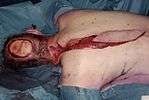
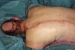
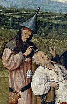
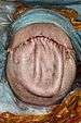
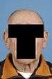
If the periosteum or underlying muscles (frontalis, occipitalis, temporalis) are intact, secondary closure by granulation is possible. Before surgical intervention this was the only option available, but aesthetically it nowadays would not be acceptable. The picture of Robert McGee illustrates this. A better option is the use of split-thickness or full-thickness skin grafts, which is quicker and gives more pleasing aesthetic results. When bulk is needed for a better contour a free flap is used, or as shown in the pictures, a regional flap.[8]
Skin reconstruction
Local reconstruction

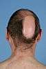
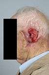
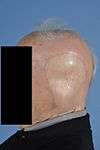
If the skin defect does not exceed 3 cm in diameter, it can be closed primarily. If this is not possible without tension, the surrounding loose connective tissue can be undermined to attain more mobility.[9] Different kinds of transpositions to close the defect with adjacent skin are possible: V-Y, Z, pinwheel flaps, advancement flaps, Orticochea flaps and rotation flaps. All these transpositions generate tension of the skin and may distort hair lines. A combination of the Orticochea and rotation flaps is illustrated by the pictures about the forehead defect. Another option is secondary healing, but this is aesthetically inferior to primary closure in hair bearing areas because of the resulting alopecia. If the scalp cannot be closed primarily and local reconstruction with hair bearing skin is needed because of hair lines, tissue expansion of hair bearing skin may be possible. The expansion process is uncomfortable on the short term, but long-term results are good.
Regional reconstruction
If local reconstruction is not possible due to lack of local tissue, regional reconstruction is the next rung on the reconstructive ladder. This includes pedicled flaps as the trapezius or supraclavicular flap or tissue expansion of nearby regions. Alternatively, the Crane principle, as described by Millard in 1969, can be used. A healthy part is used to resurface the defect and when this flap takes, the skin is returned to its original site leaving the subcutaneous tissue on the defect, which then needs a split skin graft.[1]
Skin grafts
If only skin is missing and underlying galea, muscle or connective tissue are intact, a skin graft can be used. A skin graft needs healthy, vascularised tissue beneath it to take, otherwise it will become necrotic.
Free flap
Free flaps are usually the best solution for reconstruction of large defects that cannot be closed locally and that have unfavourable wound conditions as severe infection, exposed sinuses, dura or brain tissue, CSF leakage or radiation damage. This method is the most complex of the reconstructive ladder. In scalp reconstruction free flaps have a great benefit because they are completely detached from their original location ("donor site") before transferral to the scalp which makes the inset easier compared to pedicled flaps. Another advantage is that free flaps provide a more robust vascular supply to the wound compared to pedicled flaps, controlling infection and radiation induced damage. In addition, muscle or myocutaneous free flaps provide additional bulk that obliterates empty spaces (e.g. exposed sinuses) and covers dura mater defects more than all other options, reducing postoperative wound infections and CSF leakages. Disadvantages are the complexity of the operation, leading to prolonged operation times, the need for specialised personnel, and the chance for total flap necrosis due to microvascular complications. Another challenge with the use of free flaps is to achieve an aesthetically pleasing result with good color and contour match, especially if the defect is deep.[1][10]
Outcome

Because the incidence of basal cell carcinoma and squamous-cell carcinoma is rising, there will be more need for reconstructions after radical excision of these skin malignancies. Reconstructive options depend on location and size. Most of the time the problem is not solved in just one operation, because small adjustments are necessary for good aesthetic results. Also further postoperative treatment modalities as radiation therapy and chemotherapy may impact the final result.
Aesthetic and functional
Temporal and forehead defects offer a more difficult aesthetic challenge and are best covered with a thin flap, so the aesthetic unit appears equal. Although the forehead is not a highly important aesthetic unit, color mismatch and bulkiness will draw attention quickly. Free flap reconstruction of the forehead can be bulky and color match is variable and depends on the ethnic and genetic background. Some people are not satisfied with the outcome and may experience psychological problems such as low self-confidence or even depression. Sometimes a second operation is needed to improve skin color. Overgrafting the skin with skin grafts from the scalp can improve color match.
Complications
Post-operative complications can be divided into donor-site and recipient-site problems. Donor-site complications include wound infection, hematoma, and seroma. Recipient-site complications are (total or partial) flap necrosis, wound infection, dehiscence, hematoma or skin graft failure. To avoid major bleedings or sensibility disorders, the anatomy of the scalp has to be respected. It makes a great difference if the incision is made parallel to the bloodvessel or right through it. Because of the high flow, scalp injuries can lead to serious bleedings. The cut vessels have the potential to retract into the fat so it is difficult to stop the bleeder.[6]
References
- 1 2 3 Seitz, Iris A.; Gottlieb, Lawrence J. (2009). "Reconstruction of Scalp and Forehead Defects". Clinics in Plastic Surgery. 36 (3): 355–77. doi:10.1016/j.cps.2009.02.001. PMID 19505608.
- ↑ Scalp Reconstruction Procedures at eMedicine
- 1 2 3 Seery Gerard E (2002). "Surgical anatomy of the scalp". Dermatologic Surgery. 28 (7): 581–7. doi:10.1097/00042728-200207000-00010. PMID 12135510.
- 1 2 Drake et al, Gray's Anatomy for Students, 2E, Elsevier, 2009
- ↑ Frodel, John L.; Ahlstrom, Karen (2004). "Reconstruction of Complex Scalp Defects". Archives of Facial Plastic Surgery. 6 (1): 54–60. doi:10.1001/archfaci.6.1.54. PMID 14732646.
- 1 2 Leedy, Jason E.; Janis, Jeffrey E.; Rohrich, Rod J. (2005). "Reconstruction of Acquired Scalp Defects: An Algorithmic Approach". Plastic and Reconstructive Surgery. 116 (4): 54e–72e. doi:10.1097/01.prs.0000179188.25019.6c. PMID 16163072.
- 1 2 van Driel, Antoinette A.; Mureau, Marc A. M.; Goldstein, David P.; Gilbert, Ralph W.; Irish, Jonathan C.; Gullane, Patrick J.; Neligan, Peter C.; Hofer, Stefan O. P. (2010). "Aesthetic and Oncologic Outcome after Microsurgical Reconstruction of Complex Scalp and Forehead Defects after Malignant Tumor Resection: An Algorithm for Treatment". Plastic and Reconstructive Surgery. 126 (2): 460–70. doi:10.1097/PRS.0b013e3181de2260. PMID 20679830.
- ↑ Skin Grafts and Biologic Skin Substitutes at eMedicine
- ↑ TerKonda, RP; Sykes, JM (1997). "Concepts in scalp and forehead reconstruction". Otolaryngologic Clinics of North America. 30 (4): 519–39. PMID 9233857.
- ↑ Lin, Samuel; Hanasono, Matthew; Skoracki, Roman (2008). "Scalp and Calvarial Reconstruction". Seminars in Plastic Surgery. 22 (4): 281–93. doi:10.1055/s-0028-1095887. PMC 2884877
 . PMID 20567704.