ST elevation
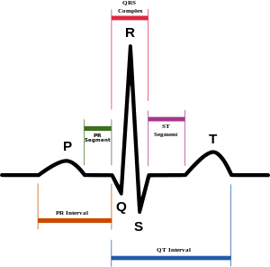

ST elevations refers to a finding on an electrocardiogram wherein the trace in the ST segment is abnormally high above the baseline.
Measurement
An ST elevation is considered significant if the vertical distance inside the ECG trace and the baseline at a point 0.24 seconds after the J-point is at least 0.9 mV (usually representing 1 mm or 1 small square) in a limb lead or 0.29 mV (2 mm or 2 small squares) in a precordial lead.[1] The baseline is either the PR interval or the TP interval.[2] This measure has a false positive rate of 15-20% (which is slightly higher in women than men) and a false negative rate of 20-30%.[3]
Physiology
The ST segment corresponds to a period of ventricular depolarization. Because of the complete depolarization of the ventricles, in theory there is no net movement of charge during the ST segment. Repolarization of the ventricle normally occurs during the T wave, however one cause of ST segment elevation is the early repolarization of the heart wall due to transmural ischemia. During transmural (subepicardial ischemia) the injured cells are "relatively" closer to the epicardial surface. In the case of depolarization, healthier cells will typically displace a larger amplitude and duration of depolarization. During systole, a ventricle with subepicardial ischemia will exhibit cells with higher amplitude of depolarization in the cardiac endocardium. Under physiological conditions the ST segment is isoelectric (i.e. same charge across the myocardium), however in some circumstances (e.g. transmural infarct) there is partial depolarization of the cardiac myocytes that have undergone ischemia. A consequence of decreased O2 perfusion will be decreased production of ATP, which is needed to operate the Na+/K+ATPase which is responsible for the final stages of myocyte repolarization (-80mV to -90mV), thus these cells will only be partially depolarized compared to surrounding healthy (non-ischemic) myocytes. The difference in membrane potential between healthy and ischemic cells causes negative charges to accumulate on their surfaces, generating a vector that points towards the normal cardiac cells (which have positive charges on their surface). This vector points away from the chest EKG leads, causing a downward deflection in the TP segment. However, since the TP segment is the baseline of the EKG, the machine corrects for this by raising TP to baseline which results in ST elevation. Also see ST depression.
Associated conditions
The exact topology and distribution of the affected areas depend on the underlying condition. Thus, ST elevation may be present on all or some leads of ECG.
It can be associated with:
- Myocardial infarction (see also ECG in myocardial infarction). ST elevation in select leads is more common with MI. ST elevation only occurs in full thickness infarction
- Prinzmetal's angina[4]
- Acute pericarditis[5][6] ST elevation in all leads is more common with acute pericarditis.
- Left ventricular aneurysm[7]
- Blunt trauma to the chest resulting in a cardiac contusion[8]
- Hyperkalemia[4]
- Acute myocarditis[4]
- Pulmonary embolism[4]
- Brugada syndrome[4]
- Hypothermia[4]
- J-point elevation[4]
- Early repolarization
See also
References
- ↑ Family Practice Notebook > ST Elevation Retrieved Nov 2010
- ↑ Khandpur, R.S. (2003). Handbook of biomedical instrumentation (2nd ed.). New Delhi: Tata McGraw-Hill. p. 255. ISBN 978-0-07-047355-3.
- ↑ Sabatine MS (2000). Pocket Medicine (이소연). Lippincott Williams & Wilkins. ISBN 0-7817-1649-7.
- 1 2 3 4 5 6 7 Thaler, Malcolm (2009). The only EKG book you'll ever need. Lippincott Williams & Wilkins. ISBN 978-1-60547-140-2.
- ↑ Tingle LE, Molina D, Calvert CW (November 2007). "Acute pericarditis". American Family Physician. 76 (10): 1509–14. PMID 18052017.
- ↑ Chew HC, Lim SH (November 2005). "Electrocardiographical case. ST elevation: is this an infarct? Pericarditis" (PDF). Singapore Medical Journal. 46 (11): 656–60. PMID 16228101.
- ↑ Victor F. Froelicher; Jonathan Myers (2006). Exercise and the heart. Elsevier Health Sciences. pp. 138–. ISBN 978-1-4160-0311-3. Retrieved 10 October 2010.
- ↑ Plautz CU, Perron AD, Brady WJ (July 2005). "Electrocardiographic ST-segment elevation in the trauma patient: acute myocardial infarction vs myocardial contusion". The American Journal of Emergency Medicine. 23 (4): 510–6. doi:10.1016/j.ajem.2004.03.014. PMID 16032622.