Right lobe of liver
| Right lobe of liver | |
|---|---|
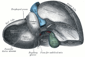 Posterior and inferior surfaces of the liver. (Right lobe labeled at upper right.) | |
 1: Right lobe of liver 2: Left lobe of liver 3: Quadrate lobe of liver 4: Round ligament of liver 5: Falciform ligament 6: Caudate lobe of liver 7: Inferior vena cava 8: Common bile duct 9: Hepatic artery 10: Portal vein 11: Cystic duct 12: Hepatic duct 13: Gallbladder | |
| Details | |
| Identifiers | |
| Latin | lobus hepatis dexter |
| TA | A05.8.01.026 |
| FMA | 13362 |
The right lobe is much larger than the left; the proportion between them being as six to one.
It occupies the right hypochondrium; on its posterior surface by the ligamentum venosum for the cranial (upper) half, and by the ligamentum teres hepatis (aka Round ligament of liver) for the caudal (under) half. The ligamentum teres hepatis turns around the inferior marging of the liver to come out ventral in the falciform ligament.
The right lobe is functionally separated from the left lobe by the middle hepatic vein. A common misconception is that the falciform ligament separates the two lobes. However, from a functional perspective (one that takes the arterial, portal venous, and systemic venous anatomy into account) the falciform ligament separates the medial and lateral segments of the left hepatic lobe.[1]
The right lobe is of a somewhat quadrilateral form. Its under and posterior surfaces being marked by three fossæ: the fossa for the portal vein, the fossa for the gall-bladder and the fossae for the inferior vena cava. These separate the right lobe in two smaller lobes on its left posterior part: the quadrate lobe and the caudate lobe.
See also
References
- ↑ Abdel-Misih, Sherif R. Z.; Bloomston, Mark (August 2010). "Liver Anatomy". Surg Clin North Am. Elsevier. 90 (4): 643–53. doi:10.1016/j.suc.2010.04.017. PMC 4038911
 .
.
This article incorporates text in the public domain from the 20th edition of Gray's Anatomy (1918)
External links
- Anatomy diagram: 12581.000-1 at Roche Lexicon - illustrated navigator, Elsevier
- Anatomy photo:37:02-0201 at the SUNY Downstate Medical Center - "Abdominal Cavity: Inspection of the Abdominal Viscera in situ"
- Anatomy photo:38:12-0204 at the SUNY Downstate Medical Center - "The Visceral Surface of the Liver"
- Cross section image: pembody/body8a - Plastination Laboratory at the Medical University of Vienna