Right coronary artery
| Right coronary artery | |
|---|---|
 Sternocostal (front) surface of heart. (Right coronary artery labeled at left.) | |
| Details | |
| Source | ascending aorta |
| Supplies | right atrium (RA), right ventricle (RV), & 25% to 35% of left ventricle. |
| Identifiers | |
| Latin | arteria coronaria dextra |
| TA | A12.2.03.101 |
| FMA | 50039 |
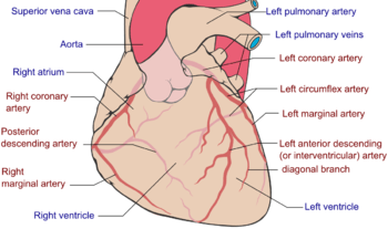
In the coronary circulation, the right coronary artery (RCA) is an artery originating above the right cusp of the aortic valve. It travels down the right atrioventricular groove, towards the crux of the heart. It branches into the posterior descending artery and the right marginal artery. Although rare, several anomalous courses of the right coronary artery have been described including origin from the left sinus of valsalva.[1]
At the origin of the RCA is the conus artery.
In addition to supplying blood to the right ventricle (RV), the RCA supplies 25% to 35% of the left ventricle (LV).
In 85% of patients (Right Dominant), the RCA gives off the posterior descending artery (PDA). In the other 15% of cases (Left Dominant), the PDA is given off by the left circumflex artery. The PDA supplies the inferior wall, ventricular septum, and the posteromedial papillary muscle.
The RCA also supplies the SA nodal artery in 60% of patients. The other 40% of the time, the SA nodal artery is supplied by the left circumflex artery.
Additional images
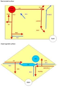 |
ARTERIES: RCA = right coronary AB = atrial branches SANB = sinuatrial nodal RMA = right marginal LCA = left coronary CB = circumflex branch LAD/AIB = anterior interventricular LMA = left marginal PIA/PDA = posterior descending MARG = left marginal AVN = atrioventricular nodal |
VEINS: SCV = small cardiac ACV = anterior cardiac AIV/GCV = great cardiac MCV = middle cardiac CS = coronary sinus |
 Right coronary artery
 Base of ventricles exposed by removal of the atria.
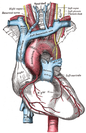 The arch of the aorta, and its branches.
 Plan of the branches.
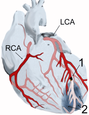 Diagram of a myocardial infarction.
 Human heart with coronary arteries
 Fetal heart - right coronary artery
References
- ↑ Angelini, P. (15 July 2014). "Novel Imaging of Coronary Artery Anomalies to Assess Their Prevalence, the Causes of Clinical Symptoms, and the Risk of Sudden Cardiac Death". Circulation: Cardiovascular Imaging. 7 (4): 747–754. doi:10.1161/CIRCIMAGING.113.000278.
External links
- Overview at Cleveland Clinic
- 00462 at CHORUS
- thoraxlesson4 at The Anatomy Lesson by Wesley Norman (Georgetown University) (heartantmajorvessels)
- Anatomy figure: 20:03-04 at Human Anatomy Online, SUNY Downstate Medical Center - "Anterior view of the heart."
- Figure of the marginal artery of the heart - merck.com.