Pyruvate kinase
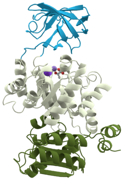 3D structure of pyruvate kinase | |||||||||
| Identifiers | |||||||||
|---|---|---|---|---|---|---|---|---|---|
| EC number | 2.7.1.40 | ||||||||
| CAS number | 9001-59-6 | ||||||||
| Databases | |||||||||
| IntEnz | IntEnz view | ||||||||
| BRENDA | BRENDA entry | ||||||||
| ExPASy | NiceZyme view | ||||||||
| KEGG | KEGG entry | ||||||||
| MetaCyc | metabolic pathway | ||||||||
| PRIAM | profile | ||||||||
| PDB structures | RCSB PDB PDBe PDBsum | ||||||||
| Gene Ontology | AmiGO / EGO | ||||||||
| |||||||||
Pyruvate kinase is the enzyme that catalyzes the final step of glycolysis. It catalyzes the transfer of a phosphate group from phosphoenolpyruvate (PEP) to adenosine diphosphate (ADP), yielding one molecule of pyruvate and one molecule of ATP.[1] Pyruvate kinase is present in four distinct, tissue-specific isozymes, each consisting of particular kinetic properties necessary to accommodate the variations in metabolic requirements of diverse tissues.
Isozymes in vertebrates
There are four isozymes of pyruvate kinase in vertebrates: L (liver), R (erythrocytes), M1(muscles, hearts and brain) and M2 (only form detectable in early fetal tissue and present in most adult tissues). R and L isozymes differ from M1 and M2 in that they are both exclusively allosterically and reversibly regulated. From a kinetic standpoint, the R and L isozymes of pyruvate kinase have two key conformation states; one with a high substrate affinity and one with a low substrate affinity. The R-state, characterized by high substrate affinity, serves as the activated form of pyruvate kinase and is stabilized by PEP and FBP, promoting the glycolytic pathway. The T-state, characterized by low substrate affinity, serves as the inactivated form of pyruvate kinase, bound and stabilized by ATP and alanine, causing phosphorylation of pyruvate kinase and the inhibition of glycolysis.[2]
Gene expression varies between the different isozymes. M1 and M2 isozymes are regulated by the gene PKM and R and L isozymes are regulated by the gene PKLR. In terms of structure, there is both a tetrameric and dimeric form of pyruvate kinase. The tetrameric form is the pyruvate kinase structure in its R-state conformation, namely with high binding affinity to PEP. In contrast, the dimeric form is its structure in T-state conformation, namely with a low binding affinity to PEP. As a result, gene expression can be regulated by converting the highly active tetrameric form of PKM2, which yields high PEP concentrations, into an inactive dimeric form, which yields a PEP concentration of nearly zero.[3]
Reaction
Glycolysis
There are two steps in the pyruvate kinase reaction in glycolysis. First, PEP transfers a phosphate group to ADP, producing ATP and the enolate of pyruvate. Secondly, a proton must be added to the enolate of pyruvate to produce the functional form of pyruvate that the cell requires.[4]

In yeast cells, the interaction of yeast pyruvate kinase (YPK) with PEP and its allosteric effector Fructose 1,6-bisphosphate (FBP,) was found to be enhanced by the presence of Mg2+. Therefore, Mg2+ was isolated as an important component in the successful catalysis of PEP into pyruvate by pyruvate kinase. Furthermore, the metal ion Mn2+ was shown to have a similar, but stronger effect on the coupling free energy of YPK than Mg2+. The binding of metal ions to the metal binding sites on pyruvate kinase enhance the rate of this glycolytic reaction.[5]
The glycolytic reaction catalyzed by pyruvate kinase is the final step of glycolysis. It is one of the three rate-affecting steps of the catabolic reaction cascade. The rate-affecting steps are the slower steps of a reaction and thus determines the rate of the overall reaction. In glycolysis, the rate-affecting steps are coupled with the hydrolysis of ATP or the phosphorylation of ADP to create the highly energetically favorable and irreversible reaction mechanism. This final step is highly regulated and deliberately irreversible because pyruvate is a crucial intermediate building block for further metabolic pathways.[6] Once pyruvate kinase synthesizes pyruvate, pyruvate either enters the TCA cycle for further production of ATP under aerobic conditions, or is reduced to lactate under anaerobic conditions. Both of these secondary metabolic pathways are essential to the function of the metabolism.
Gluconeogenesis: the reverse reaction
Pyruvate kinase also serves as a regulatory enzyme for gluconeogenesis, a biochemical pathway in which the liver generates glucose from pyruvate and other substrates. Gluconeogenesis utilizes noncarbohydrate sources to provide glucose to the brain and red blood cells in times of starvation when direct glucose reserves are exhausted.[7] During a fasting state, pyruvate carboxylase phosphorylates pyruvate kinase, inactivating the enzyme and therefore, preventing phosphoenolpyruvate from being converted into pyruvate. Instead, phosphoenolpyruvate is converted into glucose via a cascade of gluconeogenesis reactions. Although it utilizes similar enzymes, gluconeogenesis is not the reverse of glycolysis. It is instead a pathway that circumvents the irreversible steps of glycolysis. Furthermore, gluconeogenesis and glycolysis do not occur concurrently in the cell at any given moment as they are reciprocally regulated by cell signaling.[7] Once the gluconeogenesis pathway is complete, the glucose produced is expelled from the liver, proving energy for the vital tissues in the fasting state.
Regulation
Glycolysis is highly regulated at three of its catalytic steps: the phosphorylation of glucose by hexokinase, the phosphorylation of fructose-6-phosphate by phosphofructokinase, and the transfer of phosphate from PEP to ADP by pyruvate kinase. Under wild-type conditions, all three of these reactions are irreversible, have a large negative free energy and are responsible for the regulation of this pathway.[6] Pyruvate kinase activity is most broadly regulated by allosteric effectors, covalent modifiers and hormonal control. However, the most significant pyruvate kinase regulator is fructose-1,6-bisphosphate (FBP), which serves as an allosteric effector for the enzyme.
Allosteric effectors
Allosteric regulation is the binding of an effector to a site on the protein other than the active site, causing a conformational change and altering the activity of that given protein or enzyme. Pyruvate kinase has been found to be allosterically activated by FBP and allosterically inactivated by ATP and alanine.[8]
Fructose-1,6-bisphosphate
FBP is the most significant source of regulation because it comes from within the glycolysis pathway. FBP is a glycolytic intermediate produced from the phosphorylation of fructose 6-phosphate. FBP binds to the allosteric binding site on domain C of pyruvate kinase and changes the conformation of the enzyme, causing the activation of pyruvate kinase activity. As an intermediate present within the glycolytic pathway, FBP provides feedforward stimulation because the higher the concentration of FBP, the greater the allosteric activation and magnitude of pyruvate kinase activity. Pyruvate kinase is most sensitive to the effects of FBP. As a result, the remainder of the regulatory mechanisms serve as secondary modification.[9][10]
Covalent modifiers
Covalent modifiers serve as indirect regulators by controlling the phosphorylation and dephosphorylation of enzymes, resulting in the activation and inhibition of enzymatic activity. In the liver, glucagon and epinephrine serve as covalent modifiers by activating protein kinase A which in turn phosphorylates, and deactivates pyruvate kinase. In contrast, the secretion of insulin in response to blood sugar elevation activates phosphoprotein phosphatase I, causing the dephosphorylation and activation of pyruvate kinase. This regulation system is responsible for the avoidance of a futile cycle through the prevention of simultaneous activation of pyruvate kinase and enzymes that catalyze gluconeogenesis.[11]
Carbohydrate response element binding protein (ChREBP)
ChREBP is found to be an essential protein in gene transcription of the L isozyme of pyruvate kinase. The domains of ChREBP are target sites for regulation of pyruvate kinase by glucose and cAMP. Specifically, ChREBP is activated by a high concentration of glucose and inhibited by cAMP. Glucose and cAMP work in opposition with one another through covalent modifier regulation. While cAMP binds to Ser196 and Thr666 binding sites of ChREBP, causing the phosphorylation and inactivation of pyruvate kinase; glucose binds to Ser196 and Thr666 binding sites of ChREBP, causing the dephosphorylation and activation of pyruvate kinase. As a result, cAMP and excess carbohydrates are shown to play an indirect role in pyruvate kinase regulation.[12]
Hormonal control
In order to prevent a futile cycle, glycolysis and gluconeogenesis are heavily regulated in order to ensure that they are never operating in the cell at the same time. As a result, the inhibition of pyruvate kinase by glucagon, cyclic AMP and epinephrine, not only shuts down glycolysis, but also stimulates gluconeogenesis. Alternatively, insulin interferes with the effect of glucagon, cyclic AMP and epinephrine, causing pyruvate kinase to function normally and gluconeogenesis to be shut down. Furthermore, glucose was found to inhibit and disrupt gluconeogenesis, leaving pyruvate kinase activity and glycolysis unaffected. Overall, the interaction between hormones plays a key role in the functioning and regulation of glycolysis and gluconeogenesis in the cell.[13]
Inhibitory effect of metformin
Metformin, or dimethylbiguanide, is the primary treatment used for type 2 diabetes. Metaformin has been shown to indirectly affect pyruvate kinase through the inhibition of gluconeogenesis. Specifically, the addition of metformin is linked to a marked decrease in glucose flux and increase in lactate/pyruvate flux from various metabolic pathways. Although metaformin does not directly affect pyruvate kinase activity, it causes a decrease in the concentration of ATP. Due to the allosteric inhibitory effects of ATP on pyruvate kinase, a decrease in ATP results in diminished inhibition and the subsequent stimulation of pyruvate kinase. Consequently, the increase in pyruvate kinase activity directs metabolic flux through glycolysis rather than gluconeogenesis.[14]
Clinical applications
Deficiency
Genetic defects of this enzyme cause the disease known as pyruvate kinase deficiency. In this condition, a lack of pyruvate kinase slows down the process of glycolysis. This effect is especially devastating in cells that lack mitochondria, because these cells must use anaerobic glycolysis as their sole source of energy because the TCA cycle is not available. For example, red blood cells, which in a state of pyruvate kinase deficiency, rapidly become deficient in ATP and can undergo hemolysis. Therefore, pyruvate kinase deficiency can cause chronic nonspherocytic hemolytic anemia (CNSHA).[15]
PK-LR gene mutation
Pyruvate kinase deficiency is caused by an autosomal recessive trait. Mammals have two pyruvate kinase genes, PK-LR (which encodes for pyruvate kinase isozymes L and R) and PK-M (which encodes for pyruvate kinase isozyme M1), but only PKLR encodes for the red blood isozyme which effects pyruvate kinase deficiency. Over 250 PK-LR gene mutations have been identified and associated with pyruvate kinase deficiency. DNA testing has guided the discovery of the location of PKLR on chromosome 1 and the development of direct gene sequencing tests to molecularly diagnose pyruvate kinase deficiency.[16]
Applications of pyruvate kinase inhibition
Reactive Oxygen Species (ROS) Inhibition
Reactive oxygen species (ROS) are chemically reactive forms of oxygen. In human lung cells, ROS has been shown to inhibit the M2 isozyme of pyruvate kinase (PKM2). ROS achieves this inhibition by oxidizing Cys358 and inactivating PKM2. As a result of PKM2 inactivation, glucose flux is no longer converted into pyruvate, but is instead utilized in the pentose phosphate pathway, resulting in the reduction and detoxification of ROS. In this manner, the harmful effects of ROS are increased and cause greater oxidative stress on the lung cells, leading to potential tumor formation. This inhibitory mechanism is important because it may suggest that the regulatory mechanisms in PKM2 are responsible for aiding cancer cell resistance to oxidative stress and enhanced tumorigenesis.[17][18]
Phenylalanine inhibition
Phenylalanine is found to function as a competitive inhibitor of pyruvate kinase in the brain. Although the degree of phenylalanine inhibitory activity is similar in both fetal and adult cells, the enzymes in the fetal brain cells are significantly more vulnerable to inhibition than those in adult brain cells. A study of PKM2 in babies with the genetic brain disease phenylketonurics (PKU), showed elevated levels of phenylalanine and decreased effectiveness of PKM2. This inhibitory mechanism provides insight into the role of pyruvate kinase in brain cell damage.[19][20]

Alternatives
A reversible enzyme with a similar function, pyruvate phosphate dikinase (PPDK), is found in some bacteria and has been transferred to a number of anaerobic eukaryote groups (for example, Streblomastix, Giardia, Entamoeba, and Trichomonas), it seems via horizontal gene transfer on two or more occasions. In some cases, the same organism will have both pyruvate kinase and PPDK.[21]
References
- ↑ Gupta, Vibhor; Bamezai, Rameshwar N.K. (2010-11-01). "Human pyruvate kinase M2: A multifunctional protein". Protein Science. 19 (11): 2031–2044. doi:10.1002/pro.505. ISSN 1469-896X. PMC 3005776
 . PMID 20857498.
 - ↑ Muirhead, Hilary (1990-04-01). "Isoenzymes of pyruvate kinase". Biochemical Society Transactions. 18 (2): 193–196. doi:10.1042/bst0180193. ISSN 0300-5127. PMID 2379684.
- ↑ Eigenbrodt, E.; Reinacher, M.; Scheefers-Borchel, U.; Scheefers, H.; Friis, R. (1992-01-01). "Double role for pyruvate kinase type M2 in the expansion of phosphometabolite pools found in tumor cells". Critical Reviews in Oncogenesis. 3 (1-2): 91–115. ISSN 0893-9675. PMID 1532331.
- ↑ Kumar, Saroj; Barth, Andreas (2010-05-05). "Phosphoenolpyruvate and Mg2+ Binding to Pyruvate Kinase Monitored by Infrared Spectroscopy". Biophysical Journal. 98 (9): 1931–1940. doi:10.1016/j.bpj.2009.12.4335. ISSN 0006-3495. PMC 2862152. PMID 20441757.
- ↑ Bollenbach, Thomas J.; Nowak, Thomas (2001-10-01). "Kinetic Linked-Function Analysis of the Multiligand Interactions on Mg2+-Activated Yeast Pyruvate Kinase". Biochemistry. 40 (43): 13097–13106. doi:10.1021/bi010126o. ISSN 0006-2960.
- 1 2 Berg, Jeremy M.; Tymoczko, John L.; Stryer, Lubert; Berg, Jeremy M.; Tymoczko, John L.; Stryer, Lubert (2002-01-01). Biochemistry (5th ed.). W H Freeman. ISBN 0716730510.
- 1 2 Berg JM, Tymoczko JL, Stryer L. Biochemistry. 5th edition. New York: W H Freeman; 2002.
- ↑ Carbonell, Juan; Marco, Roberto; Felíu, Juan E.; Sols, Alberto (1973-08-01). "Pyruvate Kinase". European Journal of Biochemistry. 37 (1): 148–156. doi:10.1111/j.1432-1033.1973.tb02969.x. ISSN 1432-1033.
- ↑ Valentini, Giovanna; Chiarelli, Laurent; Fortin, Riccardo; Speranza, Maria L.; Galizzi, Alessandro; Mattevi, Andrea (2000-06-16). "The Allosteric Regulation of Pyruvate Kinase A SITE-DIRECTED MUTAGENESIS STUDY". Journal of Biological Chemistry. 275 (24): 18145–18152. doi:10.1074/jbc.M001870200. ISSN 0021-9258. PMID 10751408.
- ↑ Jurica, Melissa S; Mesecar, Andrew; Heath, Patrick J; Shi, Wuxian; Nowak, Thomas; Stoddard, Barry L (1998-02-15). "The allosteric regulation of pyruvate kinase by fructose-1,6-bisphosphate". Structure. 6 (2): 195–210. doi:10.1016/S0969-2126(98)00021-5.
- ↑ Birnbaum, M. J.; Fain, J. N. (1977-01-25). "Activation of protein kinase and glycogen phosphorylase in isolated rat liver cells by glucagon and catecholamines.". Journal of Biological Chemistry. 252 (2): 528–535. ISSN 0021-9258. PMID 188818.
- ↑ Kawaguchi, Takumi; Takenoshita, Makoto; Kabashima, Tsutomu; Uyeda, Kosaku (2001-11-20). "Glucose and cAMP regulate the L-type pyruvate kinase gene by phosphorylation/dephosphorylation of the carbohydrate response element binding protein". Proceedings of the National Academy of Sciences. 98 (24): 13710–13715. doi:10.1073/pnas.231370798. ISSN 0027-8424. PMC 61106. PMID 11698644.
- ↑ Feliú, J. E.; Hue, L.; Hers, H. G. (1976-08-01). "Hormonal control of pyruvate kinase activity and of gluconeogenesis in isolated hepatocytes". Proceedings of the National Academy of Sciences 73 (8): 2762–2766. ISSN 0027-8424. PMC 430732. PMID 183209.
- ↑ Argaud, Doriane; Roth, Hubert; Wiernsperger, Nicolas; Leverve, Xavier M. (1993-05-01). "Metformin decreases gluconeogenesis by enhancing the pyruvate kinase flux in isolated rat hepatocytes". European Journal of Biochemistry 213 (3): 1341–1348. doi:10.1111/j.1432-1033.1993.tb17886.x. ISSN 1432-1033.
- ↑ Grace, Rachael F.; Zanella, Alberto; Neufeld, Ellis J.; Morton, D. Holmes; Eber, Stefan; Yaish, Hassan; Glader, Bertil (2015-09-01). "Erythrocyte pyruvate kinase deficiency: 2015 status report". American Journal of Hematology. 90 (9): 825–830. doi:10.1002/ajh.24088. ISSN 1096-8652. PMID 26087744.
- ↑ Climent, Fernando; Roset, Feliu; Repiso, Ada; Pérez de la Ossa, Pablo (2009-06-01). "Red cell glycolytic enzyme disorders caused by mutations: an update". Cardiovascular & Hematological Disorders Drug Targets. 9 (2): 95–106. doi:10.2174/187152909788488636. ISSN 2212-4063. PMID 19519368.
- ↑ Anastasiou, Dimitrios; Poulogiannis, George; Asara, John M.; Boxer, Matthew B.; Jiang, Jian-kang; Shen, Min; Bellinger, Gary; Sasaki, Atsuo T.; Locasale, Jason W. (2011-12-02). "Inhibition of Pyruvate Kinase M2 by Reactive Oxygen Species Contributes to Cellular Antioxidant Responses". Science. 334 (6060): 1278–1283. doi:10.1126/science.1211485. ISSN 0036-8075. PMC 3471535. PMID 22052977.
- ↑ Christofk, Heather R.; Heiden, Matthew G. Vander; Harris, Marian H.; Ramanathan, Arvind; Gerszten, Robert E.; Wei, Ru; Fleming, Mark D.; Schreiber, Stuart L.; Cantley, Lewis C. "The M2 splice isoform of pyruvate kinase is important for cancer metabolism and tumour growth". Nature. 452 (7184): 230–233. doi:10.1038/nature06734.
- ↑ Miller, A. L.; Hawkins, R. A.; Veech, R. L. (1973-03-02). "Phenylketonuria: Phenylalanine Inhibits Brain Pyruvate Kinase in vivo". Science. 179 (4076): 904–906. doi:10.1126/science.179.4076.904. ISSN 0036-8075. PMID 4734564.
- ↑ Weber, George (1969-08-01). "Inhibition of Human Brain Pyruvate Kinase and Hexokinase by Phenylalanine and Phenylpyruvate: Possible Relevance to Phenylketonuric Brain Damage". Proceedings of the National Academy of Sciences. 63 (4): 1365–1369. doi:10.1073/pnas.63.4.1365. ISSN 0027-8424. PMC 223473. PMID 5260939.
- ↑ Liapounova, Na; Hampl, V; Gordon, Pm; Sensen, Cw; Gedamu, L; Dacks, Jb (Dec 2006), "Reconstructing the mosaic glycolytic pathway of the anaerobic eukaryote Monocercomonoides" (Free full text), Eukaryotic Cell, 5 (12): 2138–46, doi:10.1128/EC.00258-06, ISSN 1535-9778, PMC 1694820, PMID 17071828
External links
- Pyruvate kinase at the US National Library of Medicine Medical Subject Headings (MeSH)