Prophase

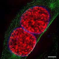
Prophase (from the Greek πρό, "before" and φάσις, "stage"), is a stage of Mitosis. This is when the chromatin condenses into two rod-shaped structures called chromosomes in which the chromatin becomes visible. This process, known as chromatin condensation, is involved with the condensin complex. Since the genetic material has been replicated in the prior interphase of the cell cycle, there are two identical copies of each chromosome in the cell. Those copies are called sister chromatids and they are attached to each other at a DNA element present on every chromosome called the centromere. Also during prophase, giemsa staining can be applied to elicit G-banding in chromosomes.
An important organelle in mitosis is the centrosome, the microtubule organizing center in metazoans. During prophase, the two centrosomes, which replicate independently of mitosis, have their microtubule-activity increased due to the recruitment of γ-tubulin. The centrosomes will be pushed apart to opposite ends of the cell nucleus by the action of molecular motors acting on the microtubules.
At the end of prophase, the tiny nucleolus within the nucleus dissolves. The nucleolus starts moving towards the nuclear membrane at the beginning of prophase, and its disintegration occurs near the nuclear envelope. Its contents are dispersed in masses.[2]
Basically, the chromatin in the nucleus coils into chromosomes. The centrosomes move to opposite poles of the cell, forming a bridge of spindle fibers. At the end of prophase, the nucleolus disperses.
In prometaphase, the next step of mitosis, the nuclear membrane breaks apart and the chromosomes are captured by the microtubules which are attached to centromeres.[3]
References
- ↑ Schermelleh, L.; Carlton, P. M.; Haase, S.; Shao, L.; Winoto, L.; Kner, P.; Burke, B.; Cardoso, M. C.; et al. (2008). "Subdiffraction Multicolor Imaging of the Nuclear Periphery with 3D Structured Illumination Microscopy". Science. 320 (5881): 1332–6. Bibcode:2008Sci...320.1332S. doi:10.1126/science.1156947. PMC 2916659
 . PMID 18535242.
 - ↑ Zeng, X.; Jiao, M.; Wang, X.; Song, Z.; Hao, S. (2001). "Electron microscopic studies on the Silver-stained Nucleolar Cycle of Physarum Polycephalum" (PDF). Acta Botanica Cinica. 43 (7): 680–5. Retrieved 24 February 2015.
- ↑ Raven, Peter H; Evert, Ray Franklin; Eichhorn, Susan E (2012). "Section 1. The Biology of the Plant Cell – 3. The Plant Cell and the Cell Cycle". Biology of plants. pp. 58–67. ISBN 978-0-7167-1007-3. LCCN 2004053303. OCLC 56051064.
External links
-
 Media related to Prophase at Wikimedia Commons