Pronator quadratus muscle
| Pronator quadratus muscle | |
|---|---|
 Anterior view of left forearm. Deep muscles. (Pronator quadratus visible at bottom-center right.) | |
| Details | |
| Origin | medial, anterior surface of the ulna |
| Insertion | lateral, anterior surface of the radius |
| Artery | anterior interosseous artery |
| Nerve | median nerve (anterior interosseous nerve) |
| Actions | pronates the forearm |
| Antagonist | Supinator muscle |
| Identifiers | |
| Latin | Musculus pronator quadratus |
| TA | A04.6.02.038 |
| FMA | 38453 |
Pronator quadratus is a square shaped muscle on the distal forearm that acts to pronate (turn so the palm faces downwards) the hand.
As it is on the anterior side of the arm, it is innervated by a branch of the median nerve, the anterior interosseous nerve (roots C8 and T1 with T1 being primary). Arterial blood comes via the interosseous artery.
Structure
Its fibres run perpendicular to the direction of the arm, running from the most distal quarter of the anterior ulna to the distal quarter of the radius. It has two heads: the superficial head originates from the anterior distal aspect of the diaphysis (shaft) of the ulna and inserts into the anterior distal diaphysis of the radius, as well as its anterior metaphysis. The deep head has the same origin, but inserts proximal to the ulnar notch.[1] It is the only muscle that attaches only to the ulna at one end and the radius at the other end.
Function
When pronator quadratus contracts, it pulls the lateral side of the radius towards the ulna, thus pronating the hand. Its deep fibers serve to keep the two bones in the forearm bound together.
Spinal Tracts
The lateral corticospinal tract is responsible for the motor pathway of the pronator quadratus. This tract begins in the precentral gyrus of the motor cortex where a signal is transmitted from the upper motor nerve through the progression tracts of the internal capsule and through the cerebral peduncles of the midbrain. It decussates in the medulla and travels down the lateral corticospinal tract in the lateral column of the spinal cord. It then decussates in the spinal cord and synapses at the anterior horn to the lower motor neurons of the skeletal muscles. The cuneate fasciculus tract is responsible for the sensation of the pronator quadratus position and movement, deep touch, visceral pain, and vibration. This tract begins in the dorsal nerve root where the signal is transmitted through the dorsal horn and up the posterior column of the spinal cord. It synapses with an interneuron in the gracile nucleus. It then decussates in the medial lemniscus of the medulla, travels through the cuneate nucleus and through the medial lemniscus of the midbrain to synapse in the thalamus. It synapses with a third order neuron and transmits the signal to the postcentral gyrus of the somesthetic cortex.
Additional Images
 Pronator quadratus muscle
 Pronator quadratus muscle
 Pronator quadratus muscle
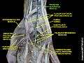 Pronator quadratus muscle
References
- ↑ Doyle, James R.; Botte, Michael J. (2003). Surgical Anatomy of the Hand and Upper Extremity. Lippincott Williams & Wilkins. pp. 129–130. ISBN 0397517254. Retrieved 1 December 2016.
External links
- 114294862 at GPnotebook
- Illustration: upper-body/pronator-quadratus from The Department of Radiology at the University of Washington
- Template:Saladin, Kenneth. Anatomy & Physiology. McGraw Hill Eduction. 2015.
- Template:Sherrington C. The Integrative Action of the Nervous System. Oxford: Oxford University Press; 1906.