Pre-eclampsia
| Pre-eclampsia | |
|---|---|
| pre-eclampsia toxaemia (PET), pre-eclampsia | |
|
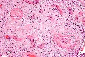
A micrograph showing hypertrophic decidual vasculopathy, a finding seen in gestational hypertension and pre-eclampsia. H&E stain. | |
| Classification and external resources | |
| Specialty | Obstetrics |
| ICD-10 | O10–O14 |
| ICD-9-CM | 642.4–642.7 |
| OMIM | 189800 |
| DiseasesDB | 10494 |
| MedlinePlus | 000898 |
| eMedicine | med/1905 ped/1885 |
| MeSH | D011225 |
| Orphanet | 275555 |
Pre-eclampsia (PE) is a disorder of pregnancy characterized by high blood pressure and often a large amount of protein in the urine.[1][2] The disorder usually occurs in the third trimester of pregnancy and worsens over time.[3][4] In severe disease there may be red blood cell breakdown, a low blood platelet count, impaired liver function, kidney dysfunction, swelling, shortness of breath due to fluid in the lungs, or visual disturbances.[3][4] Pre-eclampsia increases the risk of poor outcomes for both the mother and the baby.[4] If left untreated, it may result in seizures at which point it is known as eclampsia.[3]
Risk factors for pre-eclampsia include: obesity, prior hypertension, older age, and diabetes mellitus.[3][5] It is also more frequent in a woman's first pregnancy and if she is carrying twins.[3] The underlying mechanism involves abnormal formation of blood vessels in the placenta amongst other factors.[3] Most cases are diagnosed before delivery. Rarely, preeclampsia may begin in the period after delivery.[4] While historically both high blood pressure and protein in the urine were required to make the diagnosis, some definitions also include those with hypertension and any associated organ dysfunction.[4][6] Blood pressure is defined as high when it is greater than 140 mmHg systolic or 90 mmHg diastolic at two separate times, more than four hours apart in a woman after twenty weeks of pregnancy.[4] Preeclampsia is routinely screened for during prenatal care.[7]
Recommendations for prevention include: aspirin in those at high risk, calcium supplementation in areas with low intake, and treatment of prior hypertension with medications.[5][8] In those with preeclampsia delivery of the fetus and placenta is an effective treatment.[5] When delivery becomes recommended depends on how severe the preeclampsia and how far along in pregnancy a person is.[5] Blood pressure medication, such as labetalol and methyldopa, may be used to improve the mother's condition before delivery.[9] Magnesium sulfate may be used to prevent eclampsia in those with severe disease.[5] Bedrest and salt intake have not been found to be useful for either treatment or prevention.[4][5]
Preeclampsia affects 2–8% of pregnancies worldwide.[5] Hypertensive disorders of pregnancy (which include preeclampsia) are one of the most common causes of death due to pregnancy.[9] They resulted in 29,000 deaths in 2013 – down from 37,000 deaths in 1990.[10] Preeclampsia usually occurs after 32 weeks; however, if it occurs earlier it is associated with worse outcomes.[9] Women who have had preeclampsia are at increased risk of heart disease and stroke later in life.[7] The word eclampsia is from the Greek term for lightning.[11] The first known description of the condition was by Hippocrates in the 5th century BC.[11]
Signs and symptoms
Swelling (especially in the hands and face) was originally considered an important sign for a diagnosis of preeclampsia. However, because swelling is a common occurrence in pregnancy, its utility as a distinguishing factor in preeclampsia is not high. Pitting edema (unusual swelling, particularly of the hands, feet, or face, notable by leaving an indentation when pressed on) can be significant, and should be reported to a health care provider.
In general, none of the signs of preeclampsia are specific, and even convulsions in pregnancy are more likely to have causes other than eclampsia in modern practice. Further, a symptom such as epigastric pain may be misinterpreted as heartburn. Diagnosis, therefore, depends on finding a coincidence of several preeclamptic features, the final proof being their regression after delivery.
Causes
There is no definitive known cause of preeclampsia, though it is likely related to a number of factors. Some of these factors include:[3][7]
- Abnormal placentation (formation and development of the placenta)
- Immunologic factors
- Prior or existing maternal pathology – preeclampsia is seen more at a higher incidence in individuals with preexisting hypertension, obesity, antiphospholipid antibody syndrome, and those with history of preeclampsia
- Dietary factors, e.g. calcium supplementation in areas where dietary calcium intake is low has been shown to reduce the risk of preeclampsia[5]
- Environmental factors, e.g. air pollution[12]
Those with long term high blood pressure have a risk 7 to 8 times higher than those without.[13]
Physiologically, research has linked preeclampsia to the following physiologic changes: alterations in the interaction between the maternal immune response and the placenta, placental injury, endothelial cell injury, altered vascular reactivity, oxidative stress, imbalance among vasoactive substances, decreased intravascular volume, and disseminated intravascular coagulation.[7][14]
While the exact cause of preeclampsia remains unclear, there is strong evidence that a major cause predisposing a susceptible woman to preeclampsia is an abnormally implanted placenta.[3][7] This abnormally implanted placenta may result in poor uterine and placental perfusion, yielding a state of hypoxia and increased oxidative stress and the release of anti-angiogenic proteins along with inflammatory mediators into the maternal plasma.[7] A major consequence of this sequence of events is generalized endothelial dysfunction.[1] The abnormal implantation may stem from the maternal immune system's response to the placenta, specifically a lack of established immunological tolerance in pregnancy. Endothelial dysfunction results in hypertension and many of the other symptoms and complications associated with preclampsia.[3] Those with preeclampsia may have a lower risk of breast cancer.[15]
Risk factors
Known risk factors for preeclampsia include:[9][16]
- Nulliparity (never given birth)
- Diabetes mellitus[17]
- Kidney disease
- Chronic hypertension[17]
- Prior history of preeclampsia[17]
- Family history of preeclampsia
- Advanced maternal age (>35 years)
- Obesity[17]
- Antiphospholipid antibody syndrome[17]
- Multiple gestation[17]
- Having donated a kidney.[18]
- Having sub-clinical hypothyroidism or thyroid antibodies[19][20]
- Placental abnormalities such as placental ischemia.
Pathogenesis
Although much research into mechanism of preeclampsia has taken place, its exact pathogenesis remains uncertain. Preeclampsia is thought to result from an abnormal placenta, the removal of which ends the disease in most cases.[3] During normal pregnancy, the placenta vascularizes to allow for the exchange of water, gases, and solutes, including nutrients and wastes, between maternal and fetal circulations.[14] Abnormal development of the placenta leads to poor placental perfusion. The placenta of women with preeclampsia is abnormal and characterized by poor trophoblastic invasion.[14] It is thought that this results in oxidative stress, hypoxia, and the release of factors that promote endothelial dysfunction, inflammation, and other possible reactions.[1][14][21]
The clinical manifestations of preeclampsia are associated with general endothelial dysfunction, including vasoconstriction and end-organ ischemia.[14] Implicit in this generalized endothelial dysfunction may be an imbalance of angiogenic and anti-angiogenic factors.[3] Both circulating and placental levels of soluble fms-like tyrosine kinase-1 (sFlt-1) are higher in women with preeclampsia than in women with normal pregnancy.[14] sFlt-1 is an anti-angiogenic protein that antagonizes vascular endothelial growth factor (VEGF) and placental growth factor (PIGF), both of which are proangiogenic factors.[7] Soluble endoglin (sEng) has also been shown to be elevated in women with preeclampsia and has anti-angiogenic properties, much like sFlt-1 does.[14]
Both sFlt-1 and sEng are upregulated in all pregnant women to some extent, supporting the idea that hypertensive disease in pregnancy is a normal pregnancy adaptation gone awry. As natural killer cells are intimately involved in placentation and placentation involves a degree of maternal immune tolerance for a foreign placenta, it is not surprising that the maternal immune system might respond more negatively to the arrival of some placentae under certain circumstances, such as a placenta which is more invasive than normal. Initial maternal rejection of the placental cytotrophoblasts may be the cause of the inadequately remodeled spiral arteries in those cases of pre-eclampsia associated with shallow implantation, leading to downstream hypoxia and the appearance of maternal symptoms in response to upregulated sFlt-1 and sEng.
Oxidative stress may also play an important part in the pathogenesis of pre-eclampsia. The main source of reactive oxygen species (ROS) is the enzyme xanthine oxidase (XO) and this enzyme mainly occurs in the liver. One hypothesis is that the increased purine catabolism from placental hypoxia results in increased ROS production in the maternal liver and release into the maternal circulation that causes endothelial cell damage.[22]
Abnormalities in the maternal immune system and insufficiency of gestational immune tolerance seem to play major roles in pre-eclampsia. One of the main differences found in pre-eclampsia is a shift toward Th1 responses and the production of IFN-γ. The origin of IFN-γ is not clearly identified and could be the natural killer cells of the uterus, the placental dendritic cells modulating responses of T helper cells, alterations in synthesis of or response to regulatory molecules, or changes in the function of regulatory T cells in pregnancy.[23] Aberrant immune responses promoting pre-eclampsia may also be due to an altered fetal allorecognition or to inflammatory triggers.[23] It has been documented that fetal cells such as fetal erythroblasts as well as cell-free fetal DNA are increased in the maternal circulation in women who develop pre-eclampsia. These findings have given rise to the hypothesis that pre-eclampsia is a disease process by which a placental lesion such as hypoxia allows increased fetal material into maternal circulation, that in turn leads to an immune response and endothelial damage, and that ultimately results in pre-eclampsia and eclampsia.
One hypothesis for vulnerability to preeclampsia is the maternal-fetal conflict between the maternal organism and fetus.[24] After the first trimester trophoblasts enter the spiral arteries of the mother to alter the spiral arteries and thereby gain more access to maternal nutrients.[24] Occasionally there is impaired trophoblast invasion that results in inadequate alterations to the uterine spiral arteries.[24] It is hypothesized that the developing embryo releases biochemical signals that result in the woman developing hypertension and pre-eclampsia so that the fetus can benefit from a greater amount of maternal circulation of nutrients due to increased blood flow to the impaired placenta.[24] This results in a conflict between maternal and fetal fitness and survival because the fetus is invested in only its survival and fitness while the mother is invested in this and subsequent pregnancies.[24]
Another evolutionary hypothesis for vulnerability to pre-eclampsia is the idea of ensuring pair-bonding between the mother and father and paternal investment in the fetus.[25] Researchers posit that preeclampsia is an adaptation for the mother to terminate investment in a fetus that might have an unavailable paternal donor, as determined by repeated semen exposure of the paternal donor to the mother.[25] Various studies have shown that women who frequently had exposure to partners’ semen before conception had a reduced risk of pre-eclampsia.[25] Also, subsequent pregnancies by the same paternal donor had a reduced risk of pre-eclampsia while subsequent pregnancies by a different paternal donor had a higher risk of developing pre-eclampsia.[25]
Diagnosis
| Preeclampsia laboratory values | |
|---|---|
| Diagnostics | |
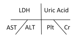 Shorthand for laboratory values commonly used in pre-eclampsia. LDH=Lactate dehydrogenase, Uric acid=Uric acid, AST=Aspartate aminotransferase, ALT=Alanine aminotransferase, Plt=Platelets, Cr=Creatinine. | |
| MeSH | D007770 |
| Reference range |
LDH: 105–333 IU/L Uric Acid: 2.4–6.0 mg/dL AST: 5–40 U/L ALT: 7–56 U/L Plt: 140–450 x 109/L Cr: 0.6–1.2 mg/dL |
| LOINC | Codes for pre-eclampsia |
Diagnostic criteria
Pre-eclampsia is diagnosed when a pregnant woman develops:[26]
- Blood pressure ≥ 140 mm Hg systolic or ≥ 90 mm Hg diastolic on two separate readings taken at least four to six hours apart after 20 weeks gestation in an individual with previously normal blood pressure.
- In a woman with essential hypertension beginning before 20 weeks gestational age, the diagnostic criteria are: an increase in systolic blood pressure (SBP) of ≥30mmHg or an increase in diastolic blood pressure (DBP) of ≥15mmHg.
- Proteinuria ≥ 0.3 grams (300 mg) or more of protein in a 24-hour urine sample or a SPOT urinary protein to creatinine ratio ≥ 0.3 or a urine dipstick reading of 1+ or greater (dipstick reading should only be used if other quantitative methods are not available).[4]
Suspicion for pre-eclampsia should be maintained in any pregnancy complicated by elevated blood pressure, even in the absence of proteinuria. Ten percent of individuals with other signs and symptoms of pre-eclampsia and 20% of individuals diagnosed with eclampsia show no evidence of proteinuria.[14] In the absence of proteinuria, the presence of new-onset hypertension (elevated blood pressure) and the new onset of one or more of the following is suggestive of the diagnosis of pre-eclampsia:[4][9]
- Evidence of kidney dysfunction (oliguria, elevated creatinine levels)
- Impaired liver function (noted by liver function tests)
- Thrombocytopenia (platelet count <100,000/microliter)
- Pulmonary edema
- Ankle edema (pitting type)
- Cerebral or visual disturbances
Pre-eclampsia is a progressive disorder and these signs of organ dysfunction are indicative of severe pre-eclampsia. A systolic blood pressure ≥160 or diastolic blood pressure ≥110 and/or proteinuria >5g in a 24-hour period is also indicative of severe pre-eclampsia.[9] Clinically, individuals with severe pre-eclampsia may also present epigastric/right upper quadrant abdominal pain, headaches, and vomiting.[9] Severe pre-eclampsia is a significant risk factor for intrauterine fetal death.
A rise in baseline blood pressure (BP) of 30 mmHg systolic or 15 mmHg diastolic, while not meeting the absolute criteria of 140/90, is important to note but is not considered diagnostic.
Predictive tests
There have been many assessments of tests aimed at predicting pre-eclampsia, though no single biomarker is likely to be sufficiently predictive of the disorder.[7] Predictive tests that have been assessed include those related to placental perfusion, vascular resistance, kidney dysfunction, endothelial dysfunction, and oxidative stress. Examples of notable tests include:
- Doppler ultrasonography of the uterine arteries to investigate for signs of inadequate placental perfusion. This test has a high negative predictive value among those individuals with a history of prior pre-eclampsia.[14]
- Elevations in serum uric acid (hyperuricemia) is used by some to "define" pre-eclampsia,[16] though it has been found to be a poor predictor of the disorder.[14] Elevated levels in the blood (hyperuricemia) are likely due to reduced uric acid clearance secondary to impaired kidney function.
- Angiogenic proteins such as vascular endothelial growth factor (VEGF) and placental growth factor (PIGF) and anti-angiogenic proteins such as soluble fms-like tyrosine kinase-1 (sFlt-1) have shown promise for potential clinical use in diagnosing pre-eclampsia, though evidence is sufficient to recommend a clinical use for these markers.[16]
- Recent studies have shown that looking for podocytes (specialized cells of the kidney) in the urine has the potential to aid in the prediction of preeclampsia. Studies have demonstrated that finding podocytes in the urine may serve as an early marker of and diagnostic test for preeclampsia.[27][28][29]
Differential diagnosis
Pre-eclampsia can mimic and be confused with many other diseases, including chronic hypertension, chronic renal disease, primary seizure disorders, gallbladder and pancreatic disease, immune or thrombotic thrombocytopenic purpura, antiphospholipid syndrome and hemolytic-uremic syndrome. It must be considered a possibility in any pregnant woman beyond 20 weeks of gestation. It is particularly difficult to diagnose when preexisting disease such as hypertension is present.[30] Women with acute fatty liver of pregnancy may also present with elevated blood pressure and protein in the urine, but differ by the extent of liver damage. Other disorders that can cause high blood pressure include thyrotoxicosis, pheochromocytoma, and drug misuse.[9]
Prevention
Preventative measures against pre-eclampsia have been heavily studied. Because the pathogensis of pre-eclampsia is not completely understood, prevention remains a complex issue. Below are some of the currently accepted recommendations.
Diet
Protein or calorie supplementation have no effect on pre-eclampsia rates, and dietary protein restriction does not appear to increase pre-eclampsia rates.[31] Further, there is no evidence that changing salt intake has an effect.[32]
Supplementation with antioxidants such as vitamin C and E has no effect on pre-eclampsia incidence,[33] nor does supplementation with vitamin D.[34] Therefore, supplementation with vitamins C, E, and D is not recommended for reducing the risk of pre-eclampsia.[34]
Calcium supplementation of at least 1 gram per day is recommended during pregnancy as it prevents preeclampsia where dietary calcium intake is low, especially for those at high risk.[34][35] Low selenium status is associated with higher incidence of pre-eclampsia.[36]
Aspirin
Taking aspirin is associated with a 1% to 5% reduction in pre-eclampsia and a 1% to 5% reduction in premature births in women at high risk.[8][37] The World Health Organization recommends low-dose aspirin for the prevention of pre-eclampsia in women at high risk and recommend it be started before 20 weeks of pregnancy.[34] The United States Preventive Services Task Force recommends a low-dose regimen for women at high risk beginning in the 12th week.[38]
Physical activity
There is insufficient evidence to recommend either exercise[39] or strict bedrest[40] as preventative measures of pre-eclampsia.
Smoking cessation
In low-risk pregnancies the association between cigarette smoking and a reduced risk of pre-eclampsia has been consistent and reproducible across epidemiologic studies. High-risk pregnancies (those with pregestational diabetes, chronic hypertension, history of pre-eclampsia in a previous pregnancy, or multifetal gestation) showed no significant protective effect. The reason for this discrepancy is not definitively known; research supports speculation that the underlying pathology increases the risk of preeclampsia to such a degree that any measurable reduction of risk due to smoking is masked.[41] However, the damaging effects of smoking on overall health and pregnancy outcomes outweighs the benefits in decreasing the incidence of preeclampsia.[7] It is recommended that smoking be stopped prior to, during and after pregnancy.[42]
Treatment
The only known definitive treatment for pre-eclampsia is delivery of the fetus and placenta. The timing of delivery should balance the desire for optimal perinatal outcomes for the fetus while reducing maternal risks.[7] The severity of disease and the maturity of the fetus are primary considerations.[43] These considerations are situation-specific and management will vary with situation, location, and institution. Treatment can range from expectant management to expedited delivery of the fetus and placenta by induction of labor or Caesarian section, in addition to pharmaceutical interventions. Important in management is the assessment of vulnerable maternal organ systems when possible, management of severe hypertension, and prevention and treatment of eclamptic seizures.[7] Separate interventions directed at the fetus may also be necessary.
Blood pressure
The World Health Organization recommends that women with severe hypertension during pregnancy should receive treatment with anti-hypertensive agents.[5] Severe hypertension is generally considered systolic BP of at least 160 or diastolic BP of at least 110.[4] Evidence does not support the use of one anti-hypertensive over another.[7] The choice of which agent to use should be based on the prescribing clinician's experience with a particular agent, its cost, and its availability.[5] Diuretics are not recommended for prevention of preeclampsia and its complications.[5] Labetolol, Hydralazine and Nifedipine are commonly used antihypertensive agents for hypertension in pregnancy.[9] ACE inhibitors and angiotensin receptor blockers are contraindicated as they affect fetal development.[26]
The goal of treatment of severe hypertension in pregnancy is to prevent cardiovascular, kidney, and cerebrovascular complications.[4] The target blood pressure has been proposed to be 140–160 mmHg systolic and 90–105 mmHg diastolic, although values are variable.[44]
Prevention of eclampsia
The intrapartum and postpartum administration of magnesium sulfate is recommended in severe pre-eclampsia for the prevention of eclampsia.[5][7] Further, magnesium sulfate is recommended for the treatment of eclampsia over other anticonvulsants.[5] Magnesium sulfate acts by interacting with NMDA receptors.[26]
Epidemiology
Pre-eclampsia affects approximately 2–8% of all pregnancies worldwide,[1][3][45] The incidence of pre-eclampsia has risen in the USA since the 1990s, possibly as a result of increased prevalence of predisposing disorders, such as chronic hypertension, diabetes, and obesity.[7]
Pre-eclampsia is one of the leading causes of maternal and perinatal morbidity and mortality worldwide.[1] Nearly one tenth of all maternal deaths in Africa and Asia and one quarter in Latin America are associated with hypertensive diseases in pregnancy, a category that encompasses pre-eclampsia.[5]
Pre-eclampsia is much more common in women who are pregnant for the first time.[46] Women who have previously been diagnosed with pre-eclampsia are also more likely to experience preeclampsia in subsequent pregnancies.[9] Pre-eclampsia is also more common in women who have preexisting hypertension, obesity, diabetes, autoimmune diseases such as lupus, various inherited thrombophilias such as Factor V Leiden, renal disease, multiple gestation (twins or multiple birth), and advanced maternal age.[9] Women who live at high altitude are also more likely to experience pre-eclampsia.[47][48] Preeclampsia is also more common in some ethnic groups (e.g. African-Americans, Sub-Saharan Africans, Latin Americans African Caribbeans and Filipinos).[7][49] Change of paternity in a subsequent pregnancy has been implicated as affecting risk, except in those with a family history of hypertensive pregnancy[50]
Eclampsia is a major complication of pre-eclampsia. Eclampsia affects 0.56 per 1000 pregnant women in developed countries and almost 10–30 times as many women in low-income countries as in developed countries.[9]
Complications
Complications of pre-eclampsia can affect both the mother and the fetus. Acutely, pre-eclampsia can be complicated by eclampsia, the development of HELLP syndrome, hemorrhagic or ischemic stroke, liver damage and dysfunction, acute kidney injury, and acute respiratory distress syndrome (ARDS).[9][14]
Pre-eclampsia is also associated with increased frequency of Caesarian section, preterm delivery, and placental abruption. Furthermore, an elevation in blood pressure can occur in some individuals in the first week postpartum attributable to volume expansion and fluid mobilization.[14] Fetal complications include fetal growth restriction and potential fetal or perinatal death.[14]
Long-term, an individual with preeclampsia is at increased risk for recurrence of pre-eclampsia in subsequent pregnancies.
Eclampsia
Eclampsia is the development of new convulsions in a pre-eclamptic patient that may not be attributed to other cause. Eclampsia is a serious complication of pre-eclampsia and results in high rates of perinatal and maternal morbidity and mortality.[5] Warning symptoms for eclampsia in an individual with current pre-eclampsia may include headaches, visual disturbances, and right upper quadrant or epigastric abdominal pain, with headache being the most consistent symptom.[7][51] Magnesium sulfate is used to prevent convulsions in cases of severe pre-eclampsia.
HELLP Syndrome
HELLP syndrome is defined as hemolysis (microangiopathic), elevated liver enzymes (liver dysfunction), and low platelets (thrombocytopenia). This condition may occur in 10–20% of patients with severe pre-eclampsia and eclampsia[7] and is associated with increased maternal and fetal morbidity and mortality. In 50% of instances, HELLP syndrome develops preterm, while 20% of cases develop in late gestation and 30% during the post-partum period.[9]
Long term
There is also an increased risk for cardiovascular complications, including hypertension and ischemic heart disease, and kidney disease.[14] Other risks include stroke and venous thromboembolism.[52][53] It seems pre-eclampsia does not increase the risk of cancer.[52]
History
The word eclampsia is from the Greek term for lightning.[11] The first known description of the condition was by Hippocrates in the 5th century BC.[11]
An outdated medical term for pre-eclampsia is toxemia of pregnancy, a term that originated in the mistaken belief that the condition was caused by toxins.[54]
Research
Some studies have suggested the importance of a woman's immunological tolerance to her baby's father, as the baby and father share genetics. There is tentative evidence that ongoing exposure either by vaginal or oral sex to the same semen that resulted in the pregnancy decreases the risk of pre-eclampsia.[55] As one early study described, "although pre-eclampsia is a disease of first pregnancies, the protective effect of multiparity is lost with change of partner".[56] The study also concluded that although women with changing partners are strongly advised to use condoms to prevent sexually transmitted diseases, "a certain period of sperm exposure within a stable relation, when pregnancy is aimed for, is associated with protection against pre-eclampsia".[56]
Several other studies have since investigated the decreased incidence of pre-eclampsia in women who had received blood transfusions from their partner, those with long preceding histories of sex without barrier contraceptives, and in women who had been regularly performing oral sex.[57]
Having already noted the importance of a woman's immunological tolerance to her baby's paternal genes, several Dutch reproductive biologists decided to take their research a step further. Consistent with the fact that human immune systems tolerate things better when they enter the body via the mouth, the Dutch researchers conducted a series of studies that confirmed a surprisingly strong correlation between a diminished incidence of pre-eclampsia and a woman's practice of oral sex, and noted that the protective effects were strongest if she swallowed her partner's semen.[57][58] A team from the University of Adelaide has also investigated to see if men who have fathered pregnancies which have ended in miscarriage or pre-eclampsia had low seminal levels of critical immune modulating factors such as TGF-Beta. The team has found that certain men, dubbed "dangerous males", are several times more likely to father pregnancies that would end in either pre-eclampsia or miscarriage.[55] Among other things, most of the "dangerous males" seemed to lack sufficient levels of the seminal immune factors necessary to induce immunological tolerance in their partners.[59]
As the theory of immune intolerance as a cause of pre-eclampsia has become accepted, women who with repeated pre-eclampsia, miscarriages, or in vitro fertilization failures could potentially be administered key immune factors such as TGF-beta along with the father's foreign proteins, possibly either orally, as a sublingual spray, or as a vaginal gel to be applied onto the vaginal wall before intercourse.[55]
References
- 1 2 3 4 5 Eiland, Elosha; Nzerue, Chike; Faulkner, Marquetta (2012). "Preeclampsia 2012". Journal of Pregnancy. 2012: 1–7. doi:10.1155/2012/586578.
- ↑ Hypertension in pregnancy. ACOG. 2013. p. 2. ISBN 9781934984284.
- 1 2 3 4 5 6 7 8 9 10 11 12 Al-Jameil, N; Aziz Khan, F; Fareed Khan, M; Tabassum, H (February 2014). "A brief overview of preeclampsia.". Journal of clinical medicine research. 6 (1): 1–7. doi:10.4021/jocmr1682w. PMID 24400024.
- 1 2 3 4 5 6 7 8 9 10 11 "Hypertension in pregnancy. Report of the American College of Obstetricians and Gynecologists' Task Force on Hypertension in Pregnancy." (PDF). Obstet Gynecol. 122 (5): 1122–31. Nov 2013. doi:10.1097/01.AOG.0000437382.03963.88. PMID 24150027.
- 1 2 3 4 5 6 7 8 9 10 11 12 13 14 15 WHO recommendations for prevention and treatment of pre-eclampsia and eclampsia. (PDF). 2011. ISBN 978-92-4-154833-5.
- ↑ Lambert, G; Brichant, JF; Hartstein, G; Bonhomme, V; Dewandre, PY (2014). "Preeclampsia: an update.". Acta Anaesthesiologica Belgica. 65 (4): 137–49. PMID 25622379.
- 1 2 3 4 5 6 7 8 9 10 11 12 13 14 15 16 17 Steegers, Eric AP; von Dadelszen, Peter; Duvekot, Johannes J; Pijnenborg, Robert (August 2010). "Pre-eclampsia". The Lancet. 376 (9741): 631–644. doi:10.1016/S0140-6736(10)60279-6. PMID 20598363.
- 1 2 Henderson, JT; Whitlock, EP; O'Connor, E; Senger, CA; Thompson, JH; Rowland, MG (May 20, 2014). "Low-dose aspirin for prevention of morbidity and mortality from preeclampsia: a systematic evidence review for the U.S. Preventive Services Task Force.". Annals of Internal Medicine. 160 (10): 695–703. doi:10.7326/M13-2844. PMID 24711050.
- 1 2 3 4 5 6 7 8 9 10 11 12 13 14 Arulkumaran, N.; Lightstone, L. (December 2013). "Severe pre-eclampsia and hypertensive crises". Best Practice & Research Clinical Obstetrics & Gynaecology. 27 (6): 877–884. doi:10.1016/j.bpobgyn.2013.07.003. PMID 23962474.
- ↑ GBD 2013 Mortality and Causes of Death, Collaborators (17 December 2014). "Global, regional, and national age-sex specific all-cause and cause-specific mortality for 240 causes of death, 1990-2013: a systematic analysis for the Global Burden of Disease Study 2013.". Lancet. 385: 117–71. doi:10.1016/S0140-6736(14)61682-2. PMC 4340604
 . PMID 25530442.
 - 1 2 3 4 Emile R. Mohler (2006). Advanced Therapy in Hypertension and Vascular Disease. PMPH-USA. pp. 407–408. ISBN 9781550093186.
- ↑ Jun Wu; Cizao Ren; Ralph J. Delfino; Judith Chung; Michelle Wilhelm; Beate Ritz (2009). "Association Between Local Traffic-Generated Air Pollution and Pre-eclampsia and Preterm Delivery in the South Coast Air Basin of California" (PDF). Environmental Health Perspectives. Archived from the original (PDF) on 2009-07-19. Retrieved 2009-07-05.
- ↑ Bramham, K; Parnell, B; Nelson-Piercy, C; Seed, PT; Poston, L; Chappell, LC (Apr 15, 2014). "Chronic hypertension and pregnancy outcomes: systematic review and meta-analysis.". BMJ (Clinical research ed.). 348: g2301. doi:10.1136/bmj.g2301. PMC 3988319. PMID 24735917.
- 1 2 3 4 5 6 7 8 9 10 11 12 13 14 Mustafa, Reem; Ahmed, Sana; Gupta, Anu; Venuto, Rocco C. (2012). "A Comprehensive Review of Hypertension in Pregnancy". Journal of Pregnancy. 2012: 1–19. doi:10.1155/2012/105918.
- ↑ Innes, KE; Byers, TE (1999). "Preeclampsia and breast cancer risk". Epidemiology. 10: 722–732. doi:10.1097/00001648-199911000-00013. JSTOR 3703514. PMID 10535787.
- 1 2 3 al.], [edited by] F. Gary Cunningham ... [et (2010). Williams obstetrics. (23rd ed.). New York: McGraw-Hill Medical. ISBN 978-0-07-149701-5.
- 1 2 3 4 5 6 Bartsch, E; Medcalf, KE; Park, AL; Ray, JG; High Risk of Pre-eclampsia Identification, Group (19 April 2016). "Clinical risk factors for pre-eclampsia determined in early pregnancy: systematic review and meta-analysis of large cohort studies.". BMJ (Clinical research ed.). 353: i1753. PMC 4837230. PMID 27094586.
- ↑ Garg, Amit X.; Nevis, Immaculate F.; McArthur, Eric; Sontrop, Jessica M.; Koval, John J.; Lam, Ngan N.; Hildebrand, Ainslie M.; Reese, Peter P.; Storsley, Leroy; Gill, John S.; Segev, Dorry L.; Habbous, Steven; Bugeja, Ann; Knoll, Greg A.; Dipchand, Christine; Monroy-Cuadros, Mauricio; Lentine, Krista L. (2014). "Gestational Hypertension and Preeclampsia in Living Kidney Donors". New England Journal of Medicine. 372: 141114133004008. doi:10.1056/NEJMoa1408932. ISSN 0028-4793.
- ↑ van den Boogaard, E; Vissenberg, R; Land, JA; et al. (2011). "Significance of (sub)clinical thyroid dysfunction and thyroid autoimmunity before conception and in early pregnancy: a systematic review". Human Reproduction Update (Review). 17 (5): 605–19. doi:10.1093/humupd/dmr024. PMID 21622978.
- ↑ Vissenberg, R; van den Boogaard, E; van Wely, M; et al. (July 2012). "Treatment of thyroid disorders before conception and in early pregnancy: a systematic review". Human Reproduction Update (Review). 18 (4): 360–73. doi:10.1093/humupd/dms007. PMID 22431565.
- ↑ Drife, James O.; Magowan, Brian. (2004). Clinical obstetrics and gynecology. Edinburgh ; New York: Saunders. pp. 367–370. ISBN 0-7020-1775-2.
- ↑ McMaster-Fay RA (2008). "Pre-eclampsia: a disease of oxidative stress resulting from the catabolism of DNA (primarily fetal) to uric acid by xanthine oxidase in the maternal liver; a hypothesis.". Bioscience Hypotheses. 1: 35–43. doi:10.1016/j.bihy.2008.01.002.
- 1 2 Laresgoiti-Servitje E, Gómez-López N, Olson DM (April 2010). "An immunological insight into the origins of pre-eclampsia". Hum Reprod Update. 16 (5): 510–24. doi:10.1093/humupd/dmq007. PMID 20388637.
- 1 2 3 4 5 Redman CW, Sargent IL (2005). "Latest Advances in Understanding Preeclampsia". Science. 308 (5728): 1592–1594. doi:10.1126/science.1111726. PMID 15947178.
- 1 2 3 4 Davis, J. A.; Gallup, G. G. J. (2006). Platek, Steven M; Shackelford, Todd K, eds. "Female Infidelity and Paternal Uncertainty (Preeclampsia and other pregnancy complications as an adaptive response to unfamiliar semen)". Evolutionary Perspectives on Male Anti-Cuckoldry Tactics: 191–204. doi:10.1017/CBO9780511617812.010. ISBN 9780511617812.
- 1 2 3 Longo, Dan L. (Dan Louis) (2012). Harrison's principles of internal medicine. New York: McGraw-Hill. pp. 55–61. ISBN 978-0-07-174889-6.
- ↑ Craici, IM; Wagner, SJ; Bailey, KR; Fitz-Gibbon, PD; Wood-Wentz, CM; Turner, ST; Hayman, SR; White, WM; Brost, BC; Rose, CH; Grande, JP; Garovic, VD (Jun 2013). "Podocyturia predates proteinuria and clinical features of preeclampsia: longitudinal prospective study.". Hypertension. 61 (6): 1289–96. doi:10.1161/HYPERTENSIONAHA.113.01115. PMC 3713793. PMID 23529165.
- ↑ "BBC News – Pre-eclampsia predicted using test during pregnancy". 2011-11-12. Retrieved 2011-11-22.
- ↑ "Mechanisms and Management of Hypertension in Pregnant Women.". Retrieved 2011-11-22.
- ↑ "Pre-eclampsia-Eclampsia". Diagnosis and management of pre-eclampsia and eclampsia. Armenian Medical Network. 2003. Retrieved 2005-11-23.
- ↑ Kramer MS, Kakuma R (2003). Kramer, Michael S, ed. "Energy and protein intake in pregnancy". Cochrane Database of Systematic Reviews (4): CD000032. doi:10.1002/14651858.CD000032. PMID 14583907.
- ↑ Duley L, Henderson-Smart D, Meher S (Oct 19, 2005). "Altered dietary salt for preventing pre-eclampsia, and its complications.". The Cochrane database of systematic reviews (4): CD005548. doi:10.1002/14651858.CD005548. PMID 16235411.
- ↑ Rumbold AR, Crowther CA, Haslam RR, Dekker GA, Robinson JS (April 2006). "Vitamins C and E and the risks of pre-eclampsia and perinatal complications". The New England Journal of Medicine. 354 (17): 1796–806. doi:10.1056/NEJMoa054186. PMID 16641396.
- 1 2 3 4 WHO recommendations for prevention and treatment of pre-eclampsia and eclampsia. 2011. ISBN 978-92-4-154833-5.
- ↑ Hofmeyr, GJ; Lawrie, TA; Atallah, AN; Duley, L; Torloni, MR (Jun 24, 2014). "Calcium supplementation during pregnancy for preventing hypertensive disorders and related problems.". The Cochrane database of systematic reviews. 6: CD001059. doi:10.1002/14651858.CD001059.pub4. PMID 24960615.
- ↑ Rayman MP, Bode P, Redman CW (November 2003). "Low selenium status is associated with the occurrence of the pregnancy disease pre-eclampsia in women from the United Kingdom". American Journal of Obstetrics and Gynecology. 189 (5): 1343–9. doi:10.1067/S0002-9378(03)00723-3. PMID 14634566.
- ↑ Duley L, Henderson-Smart DJ, Meher S, King JF (2007). "Antiplatelet agents for preventing pre-eclampsia and its complications". Cochrane Database Syst Rev (2): CD004659. doi:10.1002/14651858.CD004659.pub2. PMID 17443552.
- ↑ "Low-Dose Aspirin Use for the Prevention of Morbidity and Mortality From Preeclampsia". United States Preventive Services Task Force. Sep 2014. Retrieved 17 Sep 2014.
- ↑ Meher S, Duley L (2006). Meher, Shireen, ed. "Exercise or other physical activity for preventing pre-eclampsia and its complications". Cochrane Database of Systematic Reviews (2): CD005942. doi:10.1002/14651858.CD005942. PMID 16625645.
- ↑ Meher S, Duley L (2006). Meher, Shireen, ed. "Rest during pregnancy for preventing pre-eclampsia and its complications in women with normal blood pressure". Cochrane Database of Systematic Reviews (2): CD005939. doi:10.1002/14651858.CD005939. PMID 16625644.
- ↑ Jeyabalan A, Powers RW, Durica AR, Harger GF, Roberts JM, Ness RB (August 2008). "Cigarette Smoke Exposure and Angiogenic Factors in Pregnancy and Preeclampsia". Am. J. Hypertens. 21 (8): 943–7. doi:10.1038/ajh.2008.219. PMC 2613772. PMID 18566591.
- ↑ Whitworth, M; Dowswell, T (Oct 7, 2009). "Routine pre-pregnancy health promotion for improving pregnancy outcomes.". The Cochrane database of systematic reviews (4): CD007536. doi:10.1002/14651858.CD007536.pub2. PMID 19821424.
- ↑ Charles R.B. Beckmann ... [et al.]; American Congress of Obstetricians and Gynecologists (2010). Obstetrics and gynecology. (6th ed.). Baltimore, MD: Lippincott Williams & Wilkins. ISBN 0781788072.
- ↑ Hypertensive Disorders in Pregnancy. Version 2.0. at Nederlandse Vereniging voor Obstetrie en Gynaecologie. Date of approval: 20-05-2005
- ↑ World Health Organization (WHO). World health report 2005: make every mother and child count. Geneva: WHO; 2005, page 63
- ↑ Robbins and Cotran, Pathological Basis of Disease, 7th ed.
- ↑ http://www.nature.com/pr/journal/v54/n1/full/pr2003356a.html
- ↑ Moore, LG; Shriver, M; Bemis, L; Hickler, B; Wilson, M; Brutsaert, T; Parra, E; Vargas, E (April 2004). "Maternal adaptation to high-altitude pregnancy: an experiment of nature--a review." (PDF). Placenta. 25 Suppl A: S60–71. doi:10.1016/j.placenta.2004.01.008. PMID 15033310.
- ↑ Urquia ML, Glazier RH, Gagnon AJ, Mortensen LH, Nybo Andersen AM, Janevic T, et al. (2014). "Disparities in pre-eclampsia and eclampsia among immigrant women giving birth in six industrialised countries". BJOG. 121 (12): 1492–500. doi:10.1111/1471-0528.12758. PMC 4232918. PMID 24758368.
- ↑ Hjartardottir S, Leifsson BG, Geirsson RT, Steinthorsdottir V (2004). "Paternity change and the recurrence risk in familial hypertensive disorder in pregnancy". Hypertension in Pregnancy. 23 (2): 219–25. doi:10.1081/PRG-120037889. PMID 15369654.
- ↑ al.], [edited by] F. Gary Cunningham ... [et (2010). Williams obstetrics. (23rd ed.). New York: McGraw-Hill Medical. ISBN 978-0071497015.
- 1 2 Bellamy L, Casas JP, Hingorani AD, Williams DJ (2007). "Pre-eclampsia and risk of cardiovascular disease and cancer in later life: systematic review and meta-analysis". BMJ. 335 (7627): 974. doi:10.1136/bmj.39335.385301.BE. PMC 2072042. PMID 17975258.
- ↑ McDonald SD, Malinowski A, Zhou Q, Yusuf S, Devereaux PJ (2008). "Cardiovascular sequelae of preeclampsia/eclampsia: a systematic review and meta-analyses". Am. Heart J. 156 (5): 918–30. doi:10.1016/j.ahj.2008.06.042. PMID 19061708.
- ↑ http://www.britannica.com/EBchecked/topic/601156/toxemia-of-pregnancy
- 1 2 3 Robertson SA, Bromfield JJ, Tremellen KP (August 2003). "Seminal 'priming' for protection from pre-eclampsia-a unifying hypothesis". Journal of Reproductive Immunology. 59 (2): 253–65. doi:10.1016/S0165-0378(03)00052-4. PMID 12896827.
- 1 2 Dekker GA, Robillard PY, Hulsey TC (June 1998). "Immune maladaptation in the etiology of pre-eclampsia: a review of corroborative epidemiologic studies". Obstetrical & Gynecological Survey. 53 (6): 377–82. doi:10.1097/00006254-199806000-00023. PMID 9618714.
- 1 2 Bonney EA (December 2007). "Preeclampsia: A view through the danger model". Journal of Reproductive Immunology. 76 (1–2): 68–74. doi:10.1016/j.jri.2007.03.006. PMC 2246056. PMID 17482268.
- ↑ Mattar R, Soares RV, Daher S (February 2005). "Sexual behavior and recurrent spontaneous abortion". International Journal of Gynaecology and Obstetrics. 88 (2): 154–5. doi:10.1016/j.ijgo.2004.11.006. PMID 15694097.
- ↑ Dekker G (2002). "The partner's role in the etiology of pre-eclampsia". Journal of Reproductive Immunology. 57 (1–2): 203–15. doi:10.1016/S0165-0378(02)00039-6. PMID 12385843.
External links
- Pre-eclampsia at DMOZ
- MedlinePlus entry on high blood pressure in pregnancy
- Mayo Clinic fact sheet on pre-eclampsia